
Chest PET Scan: When to Go for It
Talk to Health Expert

Dr. Nikunj Jain
Co-Founder and HOD - Nuclear Medicine ,MBBS, DRM, DNB, FEBNM, FANMB, Dip. CBNC
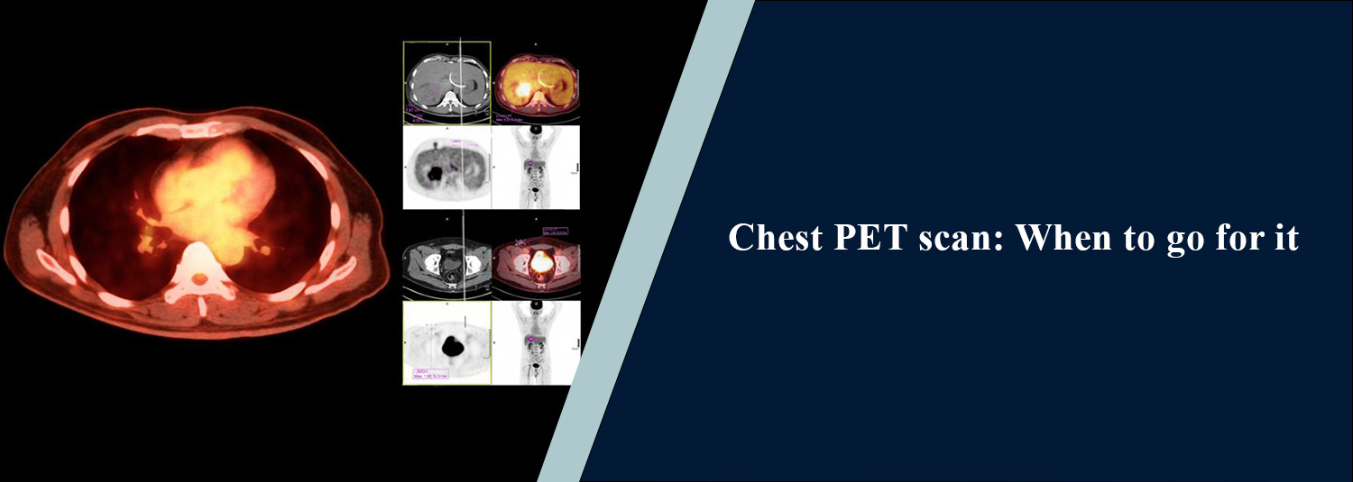
A chest PET scan is an advanced imaging
test that delivers information regarding both the structure and metabolic
activity of tissues, generally done as an integrated PET-CT scan. It can be
helpful for doctors' further evaluation/staging of selected abnormalities
already identified on CT or other imaging, if a person has a current case
of lung cancer, or if any cancer has spread to the lymph nodes or other parts
of their body.
PET-CT is not usually the first imaging
test for an undiagnosed lung abnormality. Chest computed tomography (CT) is
often used to assess the extent of pulmonary disease and lung nodules, and
PET-CT may be helpful if further information is desirable regarding the
metabolic activity or extent of disease. In Molecular Diagnostics and Therapy,
PET-CT imaging can be employed to assist with the assessment and staging of a
selected range of cancers and other disorders as needed in a clinical setting.
What Is a Chest
PET-CT Scan?
A chest PET-CT is a combination of two
imaging technologies, Positron Emission Tomography (PET) and Computed
Tomography (CT) scan. A small amount of radioactive tracer is injected into a
vein, and PET is used to gather information from metabolic activity.
Fluorodeoxyglucose (FDG) is a substance
related to the sugar glucose that is used most often in PET scans for cancer.
The CT basically gives doctors a lot of fine detail on the anatomy; they can
see where the increased and decreased tracer uptake is in the structure. When
combined, PET and CT can give complementary information that may assist a
doctor in assessing a suspected and/or known cancer.
When Is a Chest PET
Scan Recommended?
When considering previous test results
and medical history, several reasons for a chest PET scan may merit
consideration.
Evaluation of a
Suspicious Lung Nodule
A lung nodule can be found during a
chest X-ray or CT scan. Many pulmonary nodules are benign, while some are
cancerous. FDG-PET-CT could be considered in the further evaluation of some
indeterminate nodules, especially those sized and clinically appropriate to be
evaluated with PET-CT in the first place. Decisions are made by the size and
characteristics of the nodules, age, and other risk factors of a patient.
Staging of Confirmed
Lung Cancer
A PET-CT scan for lung cancer can be
useful after a diagnosis and makes use of one of the most important features of
the scan: staging. Staging is important to see if the cancer (tumor) is
contained in the lung or has migrated to the nearby lymph nodes or other parts
of the body. PET–CT can be used to give metabolic information as well as
anatomical information, and can help direct treatment planning.
Checking for Spread
of Cancer
PET-CT can be used to help determine if
there are areas of high metabolism away from the main lung mass. Depending on
the clinical situation, this can include lymph nodes and distant sites.
Findings from PET, however, are not specific for cancer. There is also a
possibility of increased FDG uptake due to infection/inflammation; therefore,
the following studies may need to be used with CT findings to interpret the
findings on PET-CT.
Evaluating Response
to Treatment
In some cases, a PET-CT can be
performed in individuals to find out whether a known cancer has responded to
treatment or not. It can give information on changes in metabolic activity,
which may be added to changes observed by anatomical imaging.
Checking for
Recurrence
PET-CT can be ordered in other patients
with cancer who have previously received treatment when clinical or imaging
history suggests that the disease has returned. It is programmed by the kind of
cancer as well as the outcomes of other examinations and previous treatment.
Is PET-CT the Same as
a Chest CT?
No. Chest CT provides detailed
anatomical images, while PET-CT combines CT with PET to provide information
about metabolic activity. PET-CT also involves radiation from both the
radiotracer and CT component. Chest computed tomography (CT) provides detailed
anatomical images of the lungs, airways, blood vessels, and other tissues in
the chest. It is a crucial imaging exam for identifying lung nodules and
numerous other lung diseases.
These anatomical images and information
on tissue metabolism are combined in a PET CT. This may be of value when
doctors want to look at the metabolic activity of a lesion or lesions and the
extent of disease in the body if they already know or are aware of having
cancer. The two examinations thus perform different functions and cannot
substitute for each other.
Chest PET-CT
Procedure
This information helps patients
understand what they can expect from the chest PET-CT procedure.
1. Preparation
Patients are typically instructed to
fast a few hours before an FDG PET-CT and can usually have water. Preparation
may differ for different tracers and exam procedures. A blood sugar level can
be tested prior to an FDG examination as well. Patients should inform the
medical team if the patient has diabetes, medications previously been
administered, has recently suffered illness, has allergies or
pregnancy/breastfeeding.
2. Tracer Injection
A small amount of radioactive tracer is injected into a vein, usually
through an IV line.
3. Uptake Period
The patient usually rests quietly for
about 30–60 minutes while the tracer circulates and is taken up by tissues.
During this time, reducing activity and talking may be a part of the
examination procedure.
4. Scanning
The patient is placed on a scanner
table inside a PET/CT scanner that is used to move the patient through the
scanner. The patient needs to remain still during imaging.
The PET and CT parts of the examination
are done together. The scanning portion commonly takes about 30 minutes; the
total appointment is longer because of preparation and the tracer uptake
period.
What Does a PET-CT
Show in Lung Cancer?
A PET-CT can provide information about metabolic activity in a lung
lesion and other areas of the body. In lung cancer evaluation, it may help
doctors assess:
- The
primary lung lesion
- Nearby lymph nodes
- Possible spread to other parts of the body
- Metabolic activity before treatment
- Changes after treatment
Remember that PET-CT can be a useful
part of the staging and treatment plan but should not supplant tissue diagnosis
if a biopsy is needed. When tissue confirmation is required, biopsy or another
appropriate tissue-sampling method may be used to confirm the diagnosis.
Does Increased PET
Uptake Always Mean Cancer?
No, increased uptake on a PET scan does not automatically mean that a
lesion is cancerous. FDG can accumulate in areas of inflammation or infection
as well as in many cancers. Conversely, some cancers may show relatively low
FDG uptake. Therefore, PET-CT findings must be interpreted in the context of
the patient's symptoms, CT findings, medical history, and other investigations.
PET-CT Test Details
The exact PET-CT scan test details depend on the tracer and the reason
for the examination. For a commonly performed FDG PET-CT:
- Fasting
may be required for several hours.
- Blood glucose may be checked.
- A small amount of radioactive tracer is
injected through an IV.
- A resting period of about 30–60 minutes may
follow.
- The patient needs to remain still during
imaging.
- The actual scanning portion is commonly around
30 minutes.
- Drinking water after the examination may help
the tracer leave the body through urine.
The diagnostic centre provides specific preparation and aftercare
instructions.
Is a Chest PET-CT
Safe?
PET-CT involves exposure to radiation from both the radioactive tracer
and CT component. The amount is controlled according to the examination protocol,
and the test is generally performed when the expected clinical benefit
justifies the exposure. Patients should tell their healthcare team if they are
pregnant, may be pregnant, or are breastfeeding before the examination.
Conclusion
A chest PET scan can give useful
information about the metabolism of the tissues, in addition to the anatomical
information obtained from a CT scan. PET-CT can help evaluate suspicious lung
masses and assess the extent of disease, as part of the overall staging process
for diagnosed lung cancer, to help identify areas that may represent regional
or distant spread to another part of the body, or to help evaluate suspected
recurrence; distinguishing recurrence from a new primary tumour may require
correlation with other imaging and sometimes tissue sampling.
A lung cancer PET scan is typically performed in addition to other tests and procedures to help with the diagnosis and staging of the cancer. A biopsy or other tissue-sampling method may be needed to confirm cancer when tissue diagnosis is required. MolecularDiagnostics and Therapy has PET-CT that is used for appropriate clinical indications, and the protocol is chosen based on the need for the exam.
Frequently Asked Questions
In News
Get Our Mobile App
for Easy Access
Book tests, view reports, and manage your health records on the go. Experience convenient healthcare with Molecular Diagnostics and Therapy.
- ✔ Book tests & home collection
- ✔ View reports instantly
- ✔ Track health history
- ✔ Get notifications & reminders
- ✔ Easy appointment management
