CEMRI BOTH KNEES
₹16000
Get CEMRI Both Knees at Molecular Diagnostics and Therapy. an NABL & NABH Accredited Diagnostic Centre providing a wide range of advanced MRI and diagnostic imaging services. Both Knees Contrast-Enhanced MRI is a high-resolution MRI of the knee with contrast enhancement when indicated clinically, to reveal a detailed evaluation of the bones, joints, cartilage, ligaments, tendons, muscles, and soft tissues surrounding the knee.
About CEMRI BOTH KNEE
CEMRI Both Knee is used to get a detailed analysis of both knee joints. An MRI scan used with intravenous (IV) contrast dye assists with the evaluation of inflammation, abnormal tissue enhancement, infection, tumors, synovial abnormalities, and other conditions of the knee. It may be recommended when symptoms affect both knees and is done to assess joint structures.
What is CEMRI BOTH KNEE?
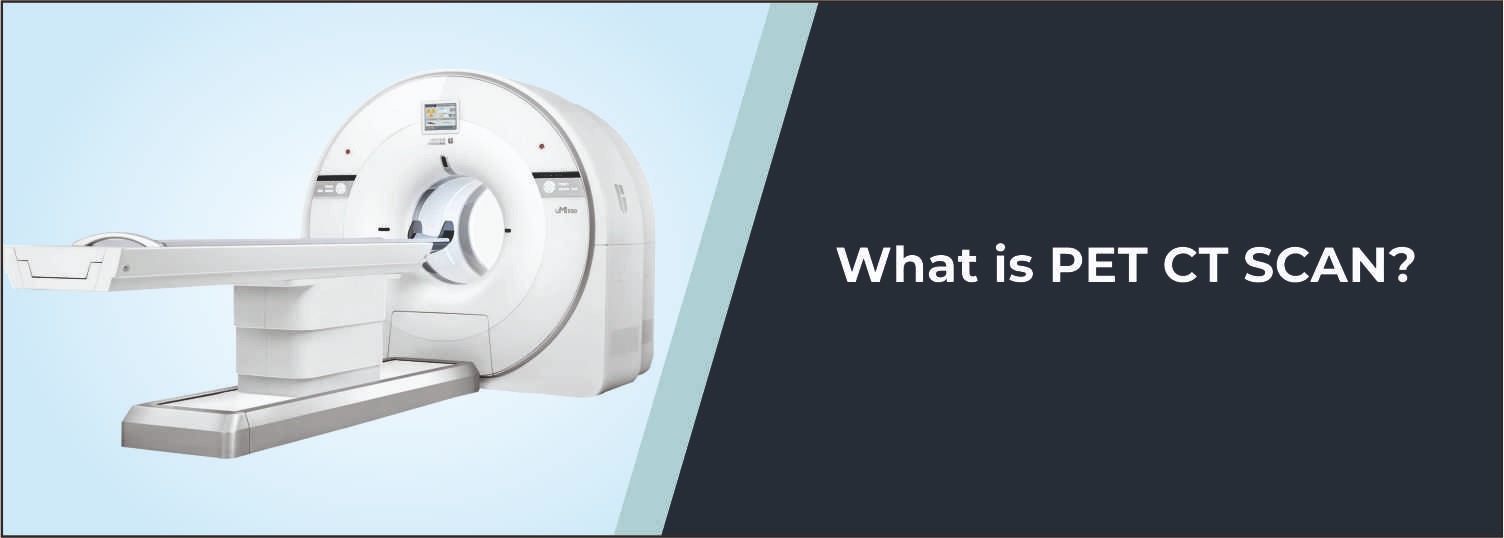
In MRI, powerful magnets and radiofrequency waves are used to construct detailed images without the use of ionizing radiation. Contrast can improve characterization of enhancement and vascularity, but non-contrast MRI can already detect many inflammatory, structural, and other abnormalities. Avoid implying that these abnormalities cannot be assessed without contrast. The scan takes detailed images of the cartilage, menisci, ligaments, tendons, surrounding soft tissue, and bone marrow.
Common Reasons for CEMRI BOTH KNEE
- Persistent or unexplained pain in both knees
- Suspected ligament or tendon abnormalities
- Evaluation of inflammatory or infective joint conditions
- Assessment of joint swelling or recurrent effusion
- Suspected bone or soft-tissue lesions
- Evaluation of synovial abnormalities
- Assessment of suspected tumors or abnormal masses
- Follow-up of known knee abnormalities
- Evaluation of postoperative or post-treatment changes
- Further assessment when previous imaging requires additional characterization
Conditions Evaluated
Ligament and Tendon Abnormalities
The scan can provide detailed visualization of the major knee ligaments and tendons, helping assess tears, sprains, degeneration, inflammation, and other structural abnormalities.
Meniscal Injuries
MRI is useful for evaluating the medial and lateral menisci for tears, degeneration, and associated changes that may contribute to pain, locking, or restricted movement.
Joint Inflammation and Synovial Abnormalities
Contrast enhancement can help characterize inflammatory changes involving the synovial lining and surrounding tissues, particularly when inflammatory or infective conditions are suspected.
Bone Marrow and Bone Lesions
CEMRI can help evaluate abnormal signals within the bones around the knee, including bone marrow changes, inflammatory processes, lesions, and selected tumor-related abnormalities.
Soft-Tissue Lesions
The scan can assess muscles, tendons, bursae, and other soft tissues around the knee and may help characterize cysts, masses, inflammatory changes, and other abnormalities.
Infection and Inflammatory Conditions
Contrast-enhanced imaging may be recommended when infection, osteomyelitis, septic arthritis, or other inflammatory processes are suspected, helping assess the extent and distribution of abnormal changes.
Benefits of CEMRI BOTH KNEES
- Detailed imaging of both knee joints
- Excellent visualization of soft tissues and joint structures
- Helps evaluate ligaments, tendons, menisci, cartilage, and bone marrow
- Contrast enhancement can improve characterization of selected abnormalities
- No ionizing radiation is used
- Useful for evaluating complex or unexplained knee symptoms
- Can assist doctors in diagnosis and treatment planning
- Allows assessment of both knees during a comprehensive imaging evaluation
Why Choose Molecular Diagnostics and Therapy?
Offering advanced radiological, nuclear medicine, and pathological services in one place, Molecular Diagnostics and Therapy is an NABL & NABH Accredited Diagnostic Centre. The advanced imaging technology, expert radiology staff, uniform imaging protocols, and patient-centred care offered by our MRI services ensure patients receive the best results. An effort is made to ensure the image quality and safety of each examination. Patients are informed in advance about the scan procedure, need for contrast, and pertinent safety precautions.
Book Your CEMRI BOTH KNEES
Get an MRI with contrast dye (dye-series MRI) for both knees at Molecular Diagnostics and Therapy. For information, bring prescriptions, MRI, CT, X-ray, and other clinical records of previous tests. It is important to let our centre know if the patient has kidney disease, a past reaction to MRI contrast, a pacemaker, surgical clips, or medical devices used for a medical purpose.
Test information: Fasting is not required.
Reporting: Within 24 hours*
- Follow the specific preparation instructions provided by the MRI team, as requirements may vary depending on the clinical indication and contrast protocol.
- Fasting may be advised before a contrast-enhanced MRI according to the centre’s protocol or the patient’s clinical condition.
- Inform the MRI team about any previous reaction to MRI contrast agents or other injectable medicines.
- Inform the medical team if you have kidney disease, reduced kidney function, or any other significant medical condition.
- Inform the MRI team about all current medicines, supplements, and ongoing treatments.
- Inform the MRI team about any pacemaker, surgical clips, artificial joints, implanted pumps, metal implants, or other medical devices.
- Tell the MRI team if you have metal fragments in your body, particularly in or near the eyes, or if you have previously worked with metal.
- Inform the medical team if you are pregnant, planning pregnancy, or there is a possibility of pregnancy.
- Bring previous MRI, CT, X-ray reports, prescriptions, and other relevant clinical records if available.
- Remove jewellery, watches, piercings, keys, mobile phones, cards, and other metallic or magnetic objects before entering the MRI examination area.
- Wear comfortable clothing without metallic components whenever possible.
- Follow all positioning and breathing instructions provided by the MRI technologist and keep both knees as still as possible during image acquisition.
- Inform the MRI team if pain, swelling, or restricted movement makes it difficult to maintain the required position.
Talk to Health Expert
Health Articles