
How Advanced PET-CT Imaging Helps in Cancer Diagnosis and Treatment Planning?
Talk to Health Expert

Dr. Nikunj Jain
Co-Founder and HOD - Nuclear Medicine ,MBBS, DRM, DNB, FEBNM, FANMB, Dip. CBNC
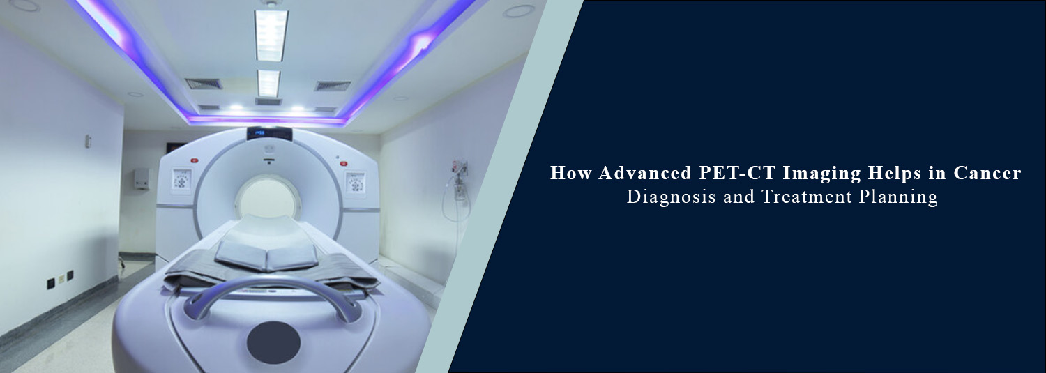
Multiple medical tests are sometimes necessary to diagnose cancer or plan the treatment. Blood tests, biopsies, and MRI and CT scans can be very helpful; however, sometimes doctors need to know not only where an area of interest is, but also how active it is. At this moment, PET-CT imaging has a significant role in the world of diagnosis. PET-CT is a combination of information from computed tomography with functional information from positron emission tomography.
The combination also assists doctors in determining the extent of disease and current cancer treatment response, and assists them in determining areas of abnormal activity and understanding what the location of the areas of abnormal activity is. Advanced cancer evaluation and treatment planning are made possible at Molecular Diagnostics and Therapy.
What Is a PET-CT Scan?
PET-CT imaging is a combination of two types of imaging at one time. The amount of the radioactive tracer used in PET reveals biological processes within the body, whereas the CT gives detailed information about the internal structures. The two pieces of information are merged together to make images that indicate what is occurring in the body and where it is occurring. Many of the PET scans done due to cancer use FDG, which is a glucose-like radioactive tracer.
What Is FDG PET-CT?
Fluorodeoxyglucose (FDG) is a PET-CT agent. It is like glucose and is absorbed to the extent that cells need to be metabolized. A few cancer cells also exhibit increased FDG uptake because of their higher glucose metabolism. This leads to the image being more noticeable in some abnormal areas.
Higher levels of FDG uptake are not necessarily associated with malignancy, however. Normal body processes also lead to increased uptake, such as in the case of infection and inflammation. The information that is seen on an FDG PET-CT is interpreted by the doctor, who also considers the patient's symptoms, medical history, biopsy results, and other investigations.
How Does PET-CT Help in Cancer Diagnosis?
A doctor may order PET-CT if they want more information about cancer they suspect or have already diagnosed, or when information from other tests isn't enough. It aids in the detection of regions with metabolic changes and can locate the region in terms of anatomy. This information is very beneficial for doctors in certain cases, when they need to evaluate whether there is a tumor or not. PET-CT should not replace biopsy in situations where tissue diagnosis is required. Rather, it supplies your diagnostic evaluation with extra information that assists with the overall evaluation.
How Can PET-CT Help Determine the Extent of Cancer?
After the diagnosis, it is important for doctors to assess whether the cancer is confined to a particular part of the body or whether it has spread elsewhere. A PET-CT scan shows parts of the body where the tracer has built up abnormally, and covers a very large area of the body. These findings, in conjunction with that of CT information, help the doctor to understand the distribution and location of disease. This information is used to help stage a cancer type, which is essential in determining the most appropriate treatment.
PET-CT and Treatment Planning
Cancer treatment can be surgical, chemotherapy, radiation therapy, immunotherapy, targeted therapy, or a combination of the above. The best treatment depends on the cancer type and size, and the condition of the patient. Along with blood markers, a PET-CT scan gives additional information that helps the doctors determine how to treat the patient's condition.
If an additional diagnosis is made elsewhere in the body, for instance, then the surgery may not be suitable, and alternative treatments might be sought. The PET-CT is thus part of a multi-step treatment planning process and not in itself a single method of treatment planning.
How Does PET-CT Help Before Surgery?
In specific cancers, PET-CT is used prior to surgery to determine the amount of cancer spread. If the imaging shows disease at some place other than the area being chosen for surgery, the plan for treating the disease might need to be changed. This assists doctors in avoiding treatment choices based on incomplete info. But, because PET-CT is not successful in reaching definitive conclusions in the field of cancer surgery, its benefit before surgery is used varies according to the type of cancer and the clinical situation of a particular patient.
Monitoring Response to Cancer Treatment
Additionally, in certain cancers, PET-CT is helpful in determining the response to treatment. If the levels of abnormal metabolic activity are reduced following treatment, it suggests that the disease is not active. Doctors also compare the results with previous scans to check if there are any changes over time. But treatment also brings about temporary changes that can alter PET results, such as inflammation. This is why it is important to follow up imaging as appropriate and to use judgment and clinical information from the treating medical team.
PET-CT for Suspected Cancer Recurrence
Symptoms, physical exams, blood tests, or other imaging changes sometimes lead to a recurrence in the mind of the doctor after chemotherapy, radiation, or other cancer treatment. PET-CT is useful in certain circumstances to find regions where there is excess metabolic activity to be further examined. It is important to remember that a positive PET-CT does not definitively diagnose recurrence. Other clinical evaluation or testing might be required to establish a cause for the abnormal finding.
How Does PET-CT Complement CT and MRI?
CT and MRI provide valuable anatomical information, but PET adds information about certain biological processes. CT is particularly useful for detailed structural imaging, while MRI can provide excellent soft-tissue contrast in many parts of the body. PET can show the distribution of a particular tracer and therefore provide information about biological activity. Using these techniques appropriately can give doctors complementary information rather than making one scan universally better than another.
The Role of Nuclear Medicine in Cancer Care
PET-CT is part of nuclear medicine, a medical specialty that uses radioactive substances for diagnosis and, in some cases, treatment. Nuclear medicine can provide information about biological processes that may not be visible through structural imaging alone. Different tracers can target different biological pathways, making molecular imaging increasingly relevant to personalised cancer care. As new tracers and technologies continue to develop, nuclear medicine may provide increasingly specific information about different diseases.
Advanced PET-CT and Future Trends in Medical Imaging
Medical imaging continues to evolve through improvements in detector technology, image reconstruction, scanning speed, and computer-based analysis. Future PET-CT systems may provide better image quality and more efficient scanning. New tracers may also allow doctors to study additional biological targets. Artificial intelligence and advanced image-analysis tools may further support specialists by helping process large amounts of imaging data and identify patterns that require closer review. These technologies are intended to support medical professionals, not replace their clinical judgement.
Can PET-CT Replace a Biopsy?
No. PET-CT cannot replace a biopsy when tissue examination is required to confirm a cancer diagnosis. A PET-CT can show areas of increased metabolic activity, but increased activity can occur for reasons other than cancer. A biopsy allows tissue to be examined directly and may be necessary for establishing a definitive diagnosis. The need for biopsy depends on the individual clinical situation.
Is PET-CT Safe?
PET-CT involves exposure to ionising radiation from both the radioactive tracer and the CT component. The amount used is determined according to the examination and clinical requirements. The decision to perform PET-CT should be based on whether the information it provides is medically useful. Patients should inform the healthcare team if they are pregnant, may be pregnant, or are breastfeeding. Patients should also follow all preparation instructions provided before the examination.
How Should Patients Prepare for an FDG PET-CT?
Preparation requirements can vary, but FDG PET-CT commonly involves specific instructions regarding food and drink before the scan. Blood glucose levels can be particularly relevant because FDG behaves similarly to glucose. Patients with diabetes may require specific preparation instructions. It is important to follow the instructions provided by the nuclear medicine team and inform them about diabetes, medications, and other relevant medical conditions before the examination.
Why Is Expert Interpretation Important?
The images produced by a PET-CT contain a large amount of information. An area of increased tracer uptake can have different causes depending on its location, intensity, pattern, and the patient's clinical history. Specialists consider these factors along with CT findings and other medical information when interpreting the examination. This is why advanced imaging technology and experienced medical professionals need to work together.
PET-CT Services at Molecular Diagnostics and Therapy
Molecular Diagnostics and Therapy offers nuclear medicine and advanced imaging services to support the evaluation and management of appropriate patients. PET-CT and FDG PET-CT can provide doctors with functional and anatomical information that may assist in cancer diagnosis, staging, treatment planning, response assessment, and evaluation of suspected recurrence. The appropriate examination is selected according to the patient's condition and the information required by the treating doctor.
Conclusion
In certain clinical scenarios, advanced PET/CT imaging has come into significance for cancer evaluation. By combining metabolic information from PET with anatomical information from CT, it can provide doctors with a more comprehensive view of disease. In many cancers, FDG PET-CT can be valuable for assessing metabolic activity, for the extent of the disease, for treatment planning, determining response to treatment, and for the evaluation of suspected recurrence.
But PET-CT is not used in place of all other diagnostic tests. However, cancer diagnosis and treatment can still benefit from other tests, including CT scans, MRI scans, blood tests, and biopsies. The future of nuclear medicine and medical imaging would see further advancement in the information that could be gathered, with the new advancements in PET-CT technology, additional tracers, and further advances in image analysis techniques. At the end of the day, the utility of any imaging modality relies upon an appropriate selection of examination for an appropriate patient and on the ability to take the entire clinical history into account with the results.
Frequently Asked Questions
In News
Get Our Mobile App
for Easy Access
Book tests, view reports, and manage your health records on the go. Experience convenient healthcare with Molecular Diagnostics and Therapy.
- ✔ Book tests & home collection
- ✔ View reports instantly
- ✔ Track health history
- ✔ Get notifications & reminders
- ✔ Easy appointment management
