
What Is a PET-CT Scan and When Do Doctors Recommend It?
Talk to Health Expert

Dr. Nikunj Jain
Co-Founder and HOD - Nuclear Medicine ,MBBS, DRM, DNB, FEBNM, FANMB, Dip. CBNC
Medical imaging has revolutionized the diagnosis and monitoring of diseases. Although not the newest imaging method, the PET CT scan is one of the most advanced available today, and combines two powerful examinations into one. While a conventional imaging test only shows the structure of the organs, a PET-CT scan gives doctors a combination of organ structure and organ function, which helps uncover abnormalities at an early stage and determine how various organs and tissues are working.
A PET-CT scan is a valuable tool in the diagnosis and treatment of a variety of disorders, such as cancer, some neurological disorders, and heart disease, because it allows medical professionals to detect changes before they are detected on regular imaging. Molecular Diagnostics and Therapy employs state-of-the-art PET CT scan imaging technology to aid in the correct diagnosis, accurate staging, treatment, and post-treatment monitoring of patients.
What Is a PET-CT Scan?
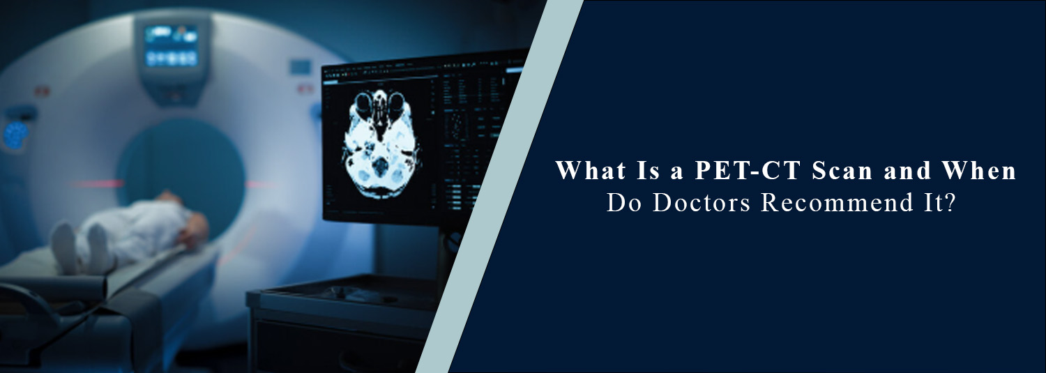
A PET-CT scan is an imaging technique used to perform a PET scan and a computed tomography (CT) scan at the same time. The PET scan detects the metabolic activity of the tissues by using a very small amount of radioactive tracer, and the CT can obtain detailed imaging information of the body's internal tissues and structures.
The combination of these two sets of images effectively allows doctors to gain a full picture, which assists with pinpointing the specific occurrence and activity of abnormalities. This hybrid can provide a more accurate diagnosis of disease than either PET or CT alone.
How Does a PET-CT Scan Work?
The radioactive tracer is given as a small shot in a vein before the scan is taken. The tracer accumulates in regions of the body with a greater metabolic rate. Following the injection, patients typically lie down for several hours so the tracer can travel around the body.
At the same time, both the PET scanner and the CT scanner can capture detailed anatomical images during the scan procedure. These images are merged together by special software to aid doctors in detecting areas of abnormal tissue growth and in finding their exact location in the body.
Why Do Doctors Recommend a PET-CT Scan?
PET-CT scans are recommended by doctors not only because they provide helpful information that can't be found with standard imaging, but also because of numerous reasons. The scan is used for the detection of diseases, their spread, the effectiveness of treatment, recovery, and disease recurrence. It also helps doctors conduct surgery or radiation therapy more accurately, or to inform treatment options for patients.
PET-CT in Cancer Diagnosis
A PET CT scan for cancer is one of the most widely used imaging techniques in the diagnosis and management of cancer. PET-CT scans can detect many types of cancer, pinpoint the exact location of a tumour, decide if cancer has spread to the lymph nodes or other organs, and determine the stage of cancer.
They also aid in determining whether there are any cancer cells that are still active or scar tissue from cancer treatment. Additionally, PET-CT imaging can also be used to check the efficacy of chemotherapy, radiation therapy, and targeted therapy if needed to adjust treatment plans in real time.
PET-CT for Heart Disease
PET–CT scans also have utility in the assessment of some cardiac diseases. This imaging test may be recommended to check the blood flow to the heart muscle, areas of damage after a heart attack, or to rule out damage to sections of the heart that could be helped by improving blood flow. This information aids cardiologists in making informed treatment decisions and achieving better outcomes for patients.
PET-CT in Neurological Disorders
PET-CT scans are used from time to time by neurologists when they are trying to assess disorders of the brain. The scan can also be used to examine abnormal brain metabolism, which may be involved with diseases like Alzheimer's disease, epilepsy, Parkinson's disease, and some forms of dementia. PET-CT may also help find the origin of the seizures before epilepsy surgery. While MRI is the imaging modality of choice for many diseases of the nervous system, the added functional data gathered by PET-CT is valuable and complements structural data.
Preparing for a PET-CT Scan
It is important to prepare the patient to obtain correct results from PET-CT. Patients may be advised to skip meals for a few hours before the examination or to drink plenty of water. Pre-test blood-sugar levels are frequently monitored before the scan, especially when diabetes exists, as high-sugar levels can have an impact on the image's quality. If a patient is pregnant, breastfeeding, has allergies, is taking medication, or has an existing medical condition, they should tell their health care provider before the examination is conducted.
What Happens During the Procedure?
Patients are given the radioactive tracer and are placed at ease for about 30 to 60 minutes as the tracer gets absorbed in the body's tissue. Typically, the scan takes about 20–45 minutes to complete. Patients then lie still on a moving motorised table, which travels through the scanner during this time. This is not painful, although it is important to be still to get good images. After the exam, most people can go back to their usual daily activities, unless their health care provider recommends otherwise.
Is a PET-CT Scan Safe?
When used for the proper medical reasons, PET-CT scans are considered to be safe. The radioactive material to be used is very small in radioactivity and is eliminated from the human body within a short period. The advantages of getting diagnostic information are usually greater than the effects of radiation. But not everyone can undergo PET-CT scans. Whether or not this imaging test is appropriate depends on your medical history and clinical condition; your doctor decides.
Benefits of PET-CT Imaging
PET-CT scanning offers several important advantages in modern medicine. It enables earlier disease detection, improves diagnostic accuracy, assists in disease staging, guides biopsy procedures, evaluates treatment response, detects recurrence, and supports personalized treatment planning. By combining functional and anatomical imaging, a full-body PET CT scan provides comprehensive information that helps doctors make more confident clinical decisions when evaluating disease throughout the body.
Conclusion
One of the most sophisticated techniques of present-day cutting-edge diagnostic imaging is PET-CT, giving information on both tissue structure and function all across the body. It can also be an important part of the diagnosis, staging, treatment planning, and follow-up in many cancers and in some heart and neurological diseases. Knowing what your physician is trying to learn from a PET–CT scan can make you feel more prepared and confident prior to the test.
Combining advanced PET-CT imaging with specialized expert staff and the latest technology, Molecular Diagnostics and Therapy provides patients with an accurate diagnosis, rigorous treatment planning, and the highest quality diagnostic testing.
Frequently Asked Questions
In News
Get Our Mobile App
for Easy Access
Book tests, view reports, and manage your health records on the go. Experience convenient healthcare with Molecular Diagnostics and Therapy.
- ✔ Book tests & home collection
- ✔ View reports instantly
- ✔ Track health history
- ✔ Get notifications & reminders
- ✔ Easy appointment management
