
Brain PET-CT Scan: Procedure, Uses, and What to Expect
Talk to Health Expert

Dr. Nikunj Jain
Co-Founder and HOD - Nuclear Medicine ,MBBS, DRM, DNB, FEBNM, FANMB, Dip. CBNC
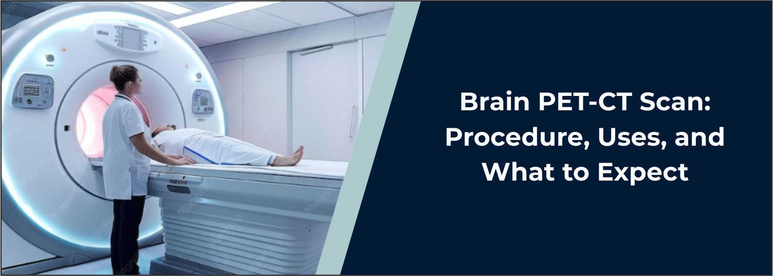
A brain PET-CT scan is an advanced
nuclear medicine imaging test that combines Positron Emission Tomography (PET)
and Computed Tomography (CT) to give information about the function as well as
the structure of the brain. While imaging tests primarily provide information
about the structure of the brain, a PET-CT can help physicians determine which
parts of the brain are consuming glucose or another particular type of tracer.
In Molecular Diagnostics and Therapy, we use powerful imaging technology
(PET-CT scan) to assess certain neurological/brain conditions.
What Is a Brain
PET-CT Scan?
Brain PET-CT is a combination of two
types of imaging. After patients receive a small tracer dose of radioactive
material, PET reveals brain regions with higher or lower glucose metabolism.
The high spatial resolution of CT helps to determine the location of these
areas. In combination, the PET and CT images can help physicians evaluate both
patterns of tracer uptake/metabolism and the actual brain structure.
Why Is a Brain PET-CT
Scan Done?
Functional brain information may be
indicated, and a PET-CT may be used to obtain an image of the brain. It can
assist the doctor in assessing the following clinical situations depending on
the type of tracer used:
- Certain
types of dementia and cognitive decline
- Alzheimer's disease and other
neurodegenerative disorders
- Epilepsy, particularly when the area
responsible for seizures needs further evaluation
- Brain tumours in selected situations
- Changes in brain metabolism associated with
certain neurological conditions
The specific indication for the test is
dependent on the exam indications, based on the patient's symptoms, medical
history, and PET tracer type employed.
How Does Brain PET-CT
Work?
A PET scan typically uses a radioactive
tracer called fluorodeoxyglucose (FDG) that is similar to glucose. Cells in the
brain normally consume glucose as their fuel; different parts of the brain may
consume glucose at different rates. Once administered, the tracer moves through
the bloodstream and is absorbed by the tissues. The PET scanner is able to
measure the radiation coming from the tracer and display the distribution of
the tracer. The CT can give detailed anatomical information, and the PET
information can be correlated with different areas of the brain.
Brain PET-CT
Procedure
The brain PET-CT procedure usually involves several steps.
1. Patient
Preparation
Preparation depends upon the type of
tracer used. In an FDG PET-CT, patients might be advised not to eat for several
hours prior to the exam. Water is permitted in general, but specific
instructions must be given. Blood glucose is also measured before the FDG scan
since inaccurate glucose levels could impact the quality of the image and how
it is interpreted.
2. Administration of
the Tracer
Trace amounts of a radioactive
substance are given in the body, typically intravenously.
3. Resting Period
Once the patient has received the
tracer, they normally lie still for a certain amount of time to allow the
tracer to distribute correctly. Patients are generally advised to rest quietly
and avoid unnecessary physical activity, talking, and major movements during
the uptake period.
4. PET-CT Imaging
The patient is positioned on the PET-CT
scanning table that is rolled through the PET-CT scanner. The patient should
remain still during image acquisition. Usually, the CT component is conducted
first or in conjunction with the combined scanning protocol, followed by PET.
How Long Does a Brain
PET-CT Take?
This scan of a brain may take a
different amount of time to complete depending on the tracer, protocol,
preparation, uptake period, and scanning requirements. An FDG scan may require
approximately 30-60 minutes for the tracer uptake to occur. The actual imaging
time varies depending on the protocol. Additional time is needed for
registration, preparation, and instructions for the patient after the scan.
What Can a Brain
PET-CT Show?
Brain PET-CT can reveal how active
various parts of the brain are. Depending upon the condition being evaluated,
metabolism changes can give additional information which complements clinical
evaluation and other imaging tests. For instance, some neurodegenerative
diseases have specific metabolic activity decreases in specific regions. During
epilepsy evaluation, areas of reduced glucose metabolism between seizures may
help identify a possible seizure-onset region. PET-CT findings should be
interpreted together with the patient's symptoms, medical history, and other
relevant investigations.
Brain PET-CT and
Alzheimer's Disease
In carefully selected individuals who
have cognitive dysfunction, PET imaging of the brain may be useful when further
information is required in the evaluation process. In some neurodegenerative
disorders, FDG PET can demonstrate a characteristic pattern of abnormal brain
glucose metabolism that can help differentiate some types of these
disorders.
Depending on clinical needs and
availability, other special PET tracers can be used to provide information on
particular biological features. PET-CT findings do not establish a diagnosis in
every patient and must be interpreted with clinical and other imaging findings.
Interpretation of the findings must be performed by an appropriately qualified
specialist, in relation to the patient's clinical picture.
Brain PET-CT for
Epilepsy
Brain PET may be used in selected
patients with drug-resistant epilepsy, particularly during presurgical
evaluation. Between seizures, areas of reduced glucose metabolism may help
identify a possible seizure-onset region. PET may be used in addition to MRI,
EEG, and other tests to make a full evaluation.
Is Brain PET-CT Safe?
PET-CT involves exposure to a relatively small amount of ionising
radiation from the radioactive tracer and CT scan. The radioactive tracer
naturally loses its radioactivity and is eliminated from the body, mainly
through urine. Patients should inform the healthcare team if they are pregnant,
may be pregnant, or are breastfeeding before the examination. The medical team
provides specific instructions based on the examination and tracer being used.
What Happens After
the Scan?
Most patients can return to their usual activities after a PET-CT unless
their healthcare provider gives different instructions. Drinking water after
the examination may help promote elimination of the tracer through urine.
Patients should follow any specific post-scan instructions provided by the
imaging centre.
Diagnostic Imaging at
Molecular Diagnostics and Therapy
At Molecular Diagnostics and
Therapy, brain PET-CT imaging is available as part of advanced diagnostic
services. The examination is performed according to the clinical requirement
and appropriate imaging protocol. The results are interpreted along with the
patient's symptoms, medical history, and other available investigations to
support clinical decision-making.
Conclusion
Brain PET-CT enables detailed
information about brain metabolism and structure to be provided by utilising
both functional PET imaging and anatomical CT imaging. It can provide
additional information during the clinical evaluation of neurological
disorders, cognitive decline, epilepsy, and selected brain tumours. The procedure
generally includes tracer administration, a resting or uptake period, and image
acquisition. Depending on the tracer, the examination may take a different
amount of time for preparation and scanning.
If your doctor has recommended a brain PET-CT scan, always follow the instructions given to you by your healthcare team to prepare for the scan, and discuss any medications, diabetes, pregnancy, or breastfeeding before having your scan. The PET-CT services at Molecular Diagnostics and Therapy are done using advanced PET imaging technology and clinical protocols for brain PET.
Frequently Asked Questions
In News
Get Our Mobile App
for Easy Access
Book tests, view reports, and manage your health records on the go. Experience convenient healthcare with Molecular Diagnostics and Therapy.
- ✔ Book tests & home collection
- ✔ View reports instantly
- ✔ Track health history
- ✔ Get notifications & reminders
- ✔ Easy appointment management
