
How Do Radiotracers Work Inside the Human Body?
Talk to Health Expert

Dr. Nikunj Jain
Co-Founder and HOD - Nuclear Medicine ,MBBS, DRM, DNB, FEBNM, FANMB, Dip. CBNC
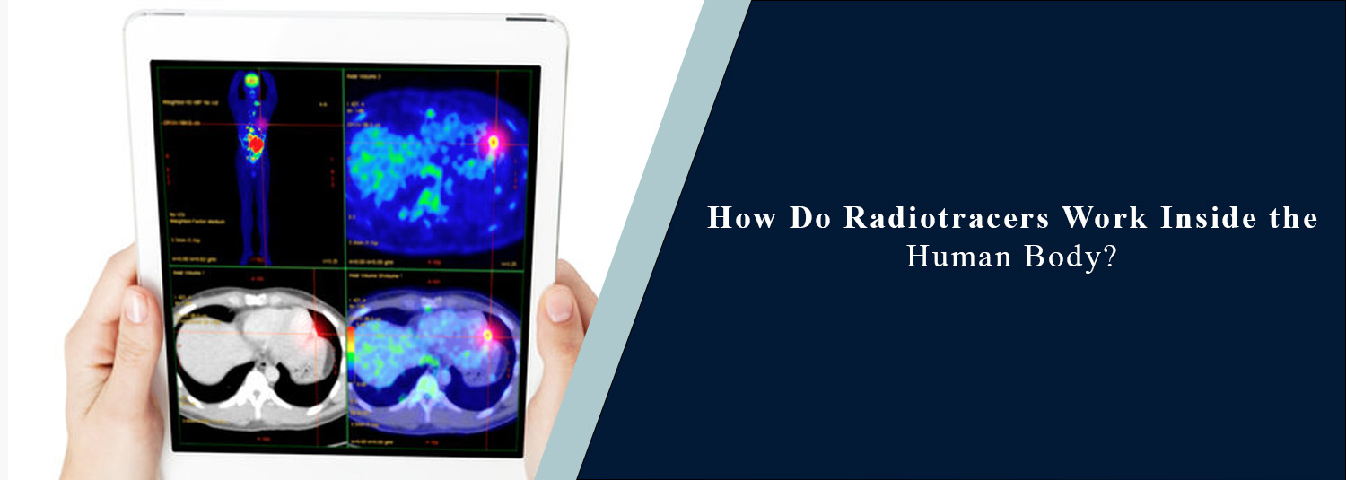
With today's modern techniques of medicine, X-ray imaging, physicians are able to examine the interior of the human body and gain knowledge of what is going on in various organs and tissues. Some scans tell primarily about the size and shape of an organ, but other scans can tell how an organ is functioning. Unsurprisingly, radiotracers are an important part of this advanced imaging technology.
In nuclear medicine, special substances known as “radiotracers” are employed to facilitate the visualization of certain activities in the body. These are administered in minute quantities and are intended to be directed to the required organ, tissue, or locality in the body. A special camera or scanner can then 'see' the signal of the radiotracer and make images.
Radiotracers are also an important part of molecular imaging, PET imaging, and other types of nuclear medicine scans. The modern nuclear medicine that we offer at Molecular Diagnostics and Therapy can give doctors a lot of information that may help them diagnose various health conditions. What Are Radiotracers? Special substances that contain trace amounts of radioactivity, such as the isotopes, are called radiotracers. They come in handy to trace certain actions within the body. Consequently, a radiotracer can be likened to a little marker. The compound is then injected into the body and moved to the targeted region to be studied by the doctors. The radioactive fraction of the tracer emits signals which can be detected by a special imaging machine.
There are multiple radiotracers for varying purposes. Some are used for the study of the heart, some for the bones, some for the kidneys, and others for the changes associated with cancer. The radionuclide selected depends on the doctor's examination procedures.
How Do Radiotracers Enter the Body? A small injection into the vein is the most common method of administration of radiotracer. In some cases, however, the radiotracer is administered orally or by other means depending upon the type of exam. Once in the body, the radiotracer is carried throughout the blood and ends up in the organs/tissues it has been designed to investigate. After receiving the tracer, the patient must wait for some time. This allows the tracer sufficient time to move to the area of interest before imaging is started. Different kinds of scans may have different lengths of time before the scan.
What Happens to a Radiotracer Inside the Body?
After administration of the radiotracer, a specific process in the body is taken. For instance, some tracers might follow cells making a more energetic effort, and other tracers might be taken up by a particular organ or follow the flow of blood. The tracer gives off a small amount of radiation. Special apparatus is developed that catches this radiation and makes an image of it.
This enables doctors to view inside the body where they may not otherwise be able to see. The radiotracer is excreted from the body after the scan. Excretion of the tracer may occur in the stool, urine, or via normal body mechanisms depending on what the tracer is.
Radiotracers and Nuclear Medicine
Radioactive materials are also used in nuclear medicine to look at the way organs and tissues function. Other imaging techniques (usually X-rays and computed tomography (CT) scans) primarily give information about the structure of the body. With nuclear medicine, you can also get other information concerning the function and activity.
A nuclear medicine scan, for instance, can provide clues to the flow of blood to the heart, the function of the kidneys, or hot spots in the body. This is possible due to the use of radiotracers, which allow the tracking of certain processes within the body.
Radiotracers in Molecular Imaging
Molecular imaging lets doctors examine the activity of cells and molecules. The cells may behave differently during a diseased state in various parts of the body. Some diseases cause cells to use energy, absorb substances, or communicate with adjoining tissues differently than they normally would.
Some of the radiotracers are made to accentuate these changes. If the tracer accumulates in a specific place, the imaging system can detect the image and display it in the scan. This can be beneficial in the process of diagnosis, treatment planning, and monitoring.
How Does PET Imaging Use Radiotracers?
While there are many applications of radiotracers, one of the most widely recognized is in PET imaging. PET scan is a test that uses a special substance called a radiotracer that is injected into the patient and moves through the body. These signals produced by the PET tracer are picked up by a PET scanner, which generates detailed images of the regions with higher and lower activity.
A glucose-based tracer is one that is commonly used for PET. Glucose is required by many cells to make energy; parts of the PET scan that have a higher amount of energy use may show greater activity than others. PET imaging is used extensively in the treatment of cancer and can sometimes be used in some heart and brain diseases.
What Is a Radiotracer Scan?
A radiotracer scan is a type of imaging test that provides information about the activity that is taking place within the body, using a radiotracer. Typically starts with giving the tracer. Following a delay, the patient is placed under or in the scanner that is suited for the kind of imaging to be administered. The imaging machine picks up the tracer and generates images that are examined by a qualified professional. Various kinds of radiotracer scans are applied to various organs and medical situations. Are Radiotracers Safe? Radiotracers are generally considered safe when used appropriately for medical imaging. Only a small amount of radioactive material is used, and the amount is carefully selected according to the examination. The radioactive material does not remain in the body permanently. The body naturally removes it over time. Patients should always inform their doctor or nuclear medicine team if they are pregnant, planning a pregnancy, or breastfeeding. This information helps the medical team decide whether the scan is suitable and whether any special precautions are required.
What Happens After a Radiotracer Scan? After most radiotracer scans, patients can return to their normal activities unless their medical team gives different instructions. Drinking enough fluids may help the body remove some tracers through urine, but patients should follow the specific instructions provided for their examination. The images are carefully reviewed by a trained nuclear medicine specialist. A report is then prepared and shared with the referring doctor.
Why Are Radiotracers Important? Radiotracers are important because they allow doctors to see processes that cannot always be seen through structural imaging alone. A CT or MRI may show what an organ looks like, while a radiotracer scan can provide information about how that organ or tissue is behaving. This additional information can be especially helpful when doctors need to understand disease activity, identify abnormal areas, plan treatment, or monitor how a patient responds to treatment.
Radiotracers in Modern Healthcare Radiotracers have become an important part of modern diagnostic medicine. They are used in many areas, including cancer imaging, heart imaging, bone scans, kidney scans, and other specialized examinations. As medical technology continues to improve, new radiotracers are being developed to target specific biological processes more accurately. This is helping doctors gain a better understanding of diseases and make more informed decisions about patient care.
Choosing the Right Nuclear Medicine Centre If your doctor has recommended a radiotracer scan, choosing an experienced nuclear medicine centre is important. At Molecular Diagnostics and Therapy, advanced nuclear medicine and molecular imaging technologies are used to support accurate diagnosis. Trained specialists carefully perform the examination, interpret the images, and prepare detailed reports for the referring doctor. The exact radiotracer and imaging method used will depend on the patient's condition and the purpose of the examination.
Conclusion Radiotracers are playing an important role in the contemporary practice of nuclear medicine. Their sole purpose is to serve as 'markers' to guide doctors into a specific activity within the body. When the particle with the radiotracer goes to the correct region, the radiotracer is detectable by special scanners that form images. Doctors can gain more information about how organs and tissues are working through molecular imaging and PET imaging, in addition to their structure.
These scans can be useful in the diagnosis, treatment planning, and follow-up of patients. In each examination, radiotracers are used in minute quantities and are carefully selected for their use. Advanced nuclear medicine technology and expert physicians assure patients of reliable, radiotracer-based imaging for future diagnosis and advice for medical care at Molecular Diagnostics and Therapy.
Frequently Asked Questions
In News
Get Our Mobile App
for Easy Access
Book tests, view reports, and manage your health records on the go. Experience convenient healthcare with Molecular Diagnostics and Therapy.
- ✔ Book tests & home collection
- ✔ View reports instantly
- ✔ Track health history
- ✔ Get notifications & reminders
- ✔ Easy appointment management
