
Lung PET-CT Scan: Uses, Procedure, Test Details and Accuracy
Talk to Health Expert

Dr. Nikunj Jain
Co-Founder and HOD - Nuclear Medicine ,MBBS, DRM, DNB, FEBNM, FANMB, Dip. CBNC
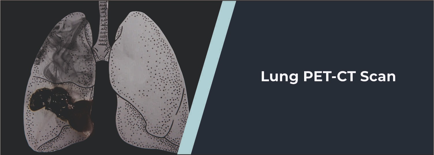
Lung cancer is one of the major types
of cancer affecting the respiratory system. Imaging tests are also used when
lung cancer is suspected or diagnosed to learn more about the size, area,
activity, and potential spread of the cancer in the lungs. Lung PET-CT Imaging
is a combination of two imaging techniques, positron emission tomography (PET)
and computed tomography (CT) imaging, performed in a single examination.
Functional and anatomical information
can be obtained from a PET- CT scan. In PET, a radiotracer is given in the
body, and it indicates regions of higher metabolic activity, whereas in CT,
detailed images of the body's structures are obtained. The combination of this
clinical appropriateness and the data can be used to assess lung cancer and make
further clinical decisions when a scan is clinically appropriate.
Molecular Diagnostics and Therapy
advanced imaging services assist physicians in the evaluation of patients who
have been recommended for a PET-CT as part of their cancer assessment.
What Is a Lung PET-CT
Scan?
A lung PET-CT scan is an imaging test
that combines PET and CT scans. A radiotracer called FDG (fluorodeoxyglucose)
is used for many cancer-related PET-CT scans. FDG is metabolised like glucose,
and tissue with higher metabolic activity might take up more. The PET part is
used to identify metabolism, and the CT part is used for visualizing the
anatomy with great detail. When both sets of information are put together,
physicians can better determine and narrow down areas to investigate further.
Why Is PET-CT Used
for Lung Cancer?
Depending on the clinical situation, a
PET-CT may be considered in individual patients who have a diagnosed or
suspected case of lung cancer. A very important function is to assess the level
of cancer once it has been diagnosed. It could aid in determining if abnormal
activity is detected in parts of the body other than the lymph nodes.
This information can be part of the
planning for staging of the cancer and may be used by the cancer team to
determine whether surgery, radiation therapy, systemic therapy, or other
treatment is appropriate. Depending on the patient's situation, PET-CT may also
be ordered to see how well the treatment is working or to check for the
presence of any recurring disease after treatment.
When May a Lung
PET-CT Be Recommended?
PET-CT is considered by doctors when
there is a lung abnormality that needs to be evaluated or when lung cancer has
been identified and the extent of disease needs to be known. It may be taken
into account in some indeterminate lung nodules, depending on the size,
appearance, clinical history, and other imaging factors. But this does not mean
it should be performed on every person who experiences respiratory symptoms or
is someone with a lung nodule. The clinical scenario is used to decide this.
How Does a Lung
PET-CT Scan Work?
A small amount of radiotracer is often
given intravenously before the scan. The patient then lies completely still
while the tracer moves around the body and gets absorbed into the tissues.
Proper preparation is important for obtaining good-quality FDG PET-CT images,
meaning that food is often eliminated for several hours prior to the test;
however, water is usually permitted. Blood sugar may also be sampled, as
elevated blood glucose could have an impact on PET findings. Depending on the
imaging centre and examination, it may be necessary to make different
preparations.
One waits a certain amount of time, and
then the patient lies on the table he or she is to be scanned on. First the CT
scan, then a PET scan. All patients will be required to lie completely still
during the scan to ensure a proper picture is captured. The scanning time is
typically 20 to 30 minutes, but will depend on the appointment time, as
preparation time will also be required, as well as the time the radiotracer
takes to circulate.
What Does a Lung
PET-CT Show?
Lung PET-CT can provide information on
both the structure and the metabolism of tissues. CT scans may be able to
detect the size and location of nodules or masses in the lungs, and give
details of structures in the proximity. The PET images can reveal regions with
higher levels of "uptake" of the tracer. This information can be
valuable in the evaluation of lung cancer to detect metabolically active disease
and to evaluate for possible metastases or involvement of lymph nodes.
But a high FDG uptake doesn't
necessarily imply cancer. A state of inflammation and infection can also
exhibit a higher metabolic rate. In the same way, there are instances where
cancer uptake of FDG may be somewhat weak. Hence, the results from a PET-CT
should be evaluated in conjunction with symptoms, past medical history, CT, and
other investigations.
Is PET-CT Accurate
for Lung Cancer?
Caution should be taken when using the
term accuracy with PET-CT. PET-CT can be useful in assessing cancer spread and
metabolic activity, but does not always confirm a diagnosis of cancer. If they
discover something suspicious using PET-CT (a procedure which uses a special
machine that captures radiation emitted by body tissues), further tests may be
performed. Tissue sampling by biopsy is also performed in a lot of suspected
cases of lung cancer to confirm the diagnosis. Biopsy might be done with
bronchoscopy or by techniques performed with computed tomography (CT) guidance,
depending on where the abnormality is located. PET-CT may thus be valuable in
the diagnosis or staging process, although results must be evaluated in
conjunction with other tests and/or clinical data.
PET-CT for Lung
Cancer Staging
The stage of lung cancer is a
measurement of the depth of disease or whether it is spread. In non-small-cell
lung cancer and in some small-cell lung cancer (SCLC) cases, PET-CT may be
useful in staging. Depending on the patient's condition, physicians might make
use of PET-CT alongside different imaging tests (including brain MRI) and/or
biopsy and lab tests. The specific tests will depend on the type of cancer
being considered and what information is available.
Can PET-CT Be Used
After Lung Cancer Treatment?
Yes, in selected situations. PET-CT may help doctors assess how a cancer
responds to treatment or evaluate suspected recurrence after treatment. Changes
in metabolic activity can sometimes provide useful information about treatment
response. However, treatment-related inflammation can also affect PET findings,
so scan results need to be interpreted in the context of the timing of
treatment and other clinical information.
Is a Lung PET-CT Scan
Safe?
PET-CT involves exposure to radiation from both the radiotracer and CT
component. The amount used for diagnostic imaging is kept as low as reasonably
appropriate for the examination, and the potential benefits are considered
against the radiation risk. The radiotracer itself is used in a small amount
and loses radioactivity over time. It is also eliminated from the body
naturally, including through urine. Drinking water after the examination may
help with clearance unless the healthcare team gives different instructions.
Patients should inform their doctor and imaging team if they are pregnant or
breastfeeding.
How to Prepare for a
Lung PET-CT Scan
Preparation instructions should always be followed as provided by the
imaging centre. For many FDG PET-CT examinations, patients are asked not to eat
for several hours before the scan and to drink water as instructed. People with
diabetes may receive specific instructions because blood glucose levels can
affect the quality and interpretation of an FDG PET scan. Patients should also
inform the medical team about their medicines, medical conditions, recent
illnesses, and any previous imaging or treatment.
Understanding Your
PET-CT Report
A PET-CT report is usually prepared by a radiologist or nuclear medicine
physician trained in interpreting these examinations. The report may describe
findings in the lungs, lymph nodes, other organs, and bones depending on the
area scanned. The report may also include an Impression or Conclusion, where
the most important findings and recommendations are summarized. If a finding is
uncertain, the report may recommend another imaging test, follow-up, or tissue
sampling. Patients should discuss their report with the treating doctor rather
than trying to interpret individual terms without clinical context.
Why Choose Molecular
Diagnostics and Therapy for PET-CT?
Molecular Diagnostics and Therapy provides advanced diagnostic services,
including PET-CT imaging, to support doctors in evaluating various conditions.
The centre uses modern imaging technology and provides PET-CT services based on
clinical requirements. For patients undergoing evaluation for lung cancer,
PET-CT findings can provide useful information about metabolic activity and the
possible extent of disease. The final interpretation should always be
considered along with the patient's clinical history, previous imaging, biopsy
findings, and other relevant investigations.
Conclusion
It can be helpful in selected patients with lung cancer for lymph node evaluation, staging, treatment-response assessment, and evaluation of suspected recurrence. PET/CT is useful, but cannot definitively diagnose lung cancer in all situations. Other diseases and inflammation may also alter PET results, and a biopsy is often needed to confirm a diagnosis of lung cancer by examining tissue. Following your doctor's advice on preparation for a PET-CT scan for lung cancer and discussing the end result with the doctor treating you is a way to ensure that the results are considered in the proper clinical context.
Frequently Asked Questions
In News
Get Our Mobile App
for Easy Access
Book tests, view reports, and manage your health records on the go. Experience convenient healthcare with Molecular Diagnostics and Therapy.
- ✔ Book tests & home collection
- ✔ View reports instantly
- ✔ Track health history
- ✔ Get notifications & reminders
- ✔ Easy appointment management
