
Pregnancy Ultrasound Schedule: Month-by-Month Guide
Talk to Health Expert

Dr. Nikunj Jain
Co-Founder and HOD - Nuclear Medicine ,MBBS, DRM, DNB, FEBNM, FANMB, Dip. CBNC
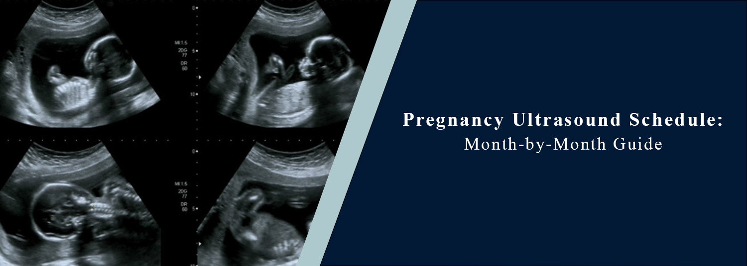
Pregnancy is a very emotional and exciting period, and to keep the mother and the developing baby healthy, frequent examinations by the doctors are necessary. One of the most important tests that is carried out in pregnancy is an ultrasound. An ultrasound during pregnancy benefits a doctor to better monitor the development of the baby, to know if the baby is in good condition, to assess if there are any complications, etc., and to provide a fetal due date.
Pregnant women are confused about the number of ultrasounds needed and when they can be done during their pregnancy. Each doctor might perform different scans at certain stages of the pregnancy, but these are the important scans that are to be expected, depending on a woman's health and medical history. There is a specific purpose and function to each scan, and there is lots of very important information to be taken from the scans regarding the baby's growth, which enables doctors to know how the baby is doing and helps restructure how the mother should eat to ensure the growth and development are optimal for the fetus.
In the modern era, monitoring of pregnancies has become easier and more accurate than ever before with modern technology in ultrasound. Molecular Diagnostics and Therapy provides advanced pregnancy ultrasound imaging and complex diagnostic services to help to ensure accurate fetal monitoring and maternal care in pregnancy.
First Month of Pregnancy
Some pregnant women do not need an ultrasound scan during the first few weeks of pregnancy unless they have signs or symptoms like bleeding, pain in the abdomen, or problems with pregnancies in the past. During a very early pregnancy, it is possible that the sac will not show up clearly. If there are concerns about an ectopic pregnancy or about the date of conception, doctors might suggest an early ultrasound.
Second Month of Pregnancy
This scan is a preliminary scan in the early stages of pregnancy, when the mother is about 6 to 8 weeks pregnant. Scanning helps to confirm whether the pregnancy is in the uterus, find the foetal heartbeat, confirm the number of foetuses, and determine the length of gestation. This move is additionally critical to not overlook ectopic and early pregnancy problems. In certain pregnancies, it may be necessary to use a transvaginal ultrasound, as this provides a better ultrasound image in early pregnancy.
Third Month of Pregnancy
Doctors generally suggest the NT scan (also known as Nuchal Translucency Scan) take place between 11 and 13 weeks. This ultrasound helps assess the baby's early development and can be used to screen for some chromosomal defects. At this time, doctors also check the baby's heartbeat rate, growth, fetal position, and early organ development. A first-trimester ultrasound is valuable for confirming the proper dating of pregnancy and the date of delivery.
Fourth Month of Pregnancy
The baby grows rapidly in the first four months. There are some women who don't need a big ultrasound at this time unless they need monitoring. If the risk of complications is high or if there are symptoms such as decreased fetal growth, pain, or bleeding, routine ultrasounds could continue. Using advanced prenatal imaging technology, Molecular Diagnostics and Therapy helps to further assess and image the fetus during and throughout pregnancies and to monitor the health of the mother.
Fifth Month of Pregnancy
During the 5th month of pregnancy, an ultrasound is one of the most important tests. Generally, doctors recommend the Anomaly Scan (also called the Level 2 Ultrasound) between 18 and 22 weeks. It helps in the evaluation of all an infant's organs and body systems, including the brain, spinal cord, heart, kidneys, arms or legs, face, stomach, and placenta. An anomaly scan detects structural anomalies, developmental abnormalities, and some congenital abnormalities. A doctor also checks the amount of amniotic fluid and fetal growth in this stage.
Sixth Month of Pregnancy
By 6 months, the ultrasound is used to confirm the progress of the pregnancy and growth of the fetus. The baby's movements, position, placenta position and size, and amniotic fluid are checked. Scans may also be required if complications have arisen during previous pregnancies, twins have been born, or the mother has diabetes and/or high blood pressure.
Seventh Month of Pregnancy
Fetal monitoring becomes more crucial in the third trimester of pregnancy. In the 7th month, an ultrasound is used to look at the body's growth rate, blood flow, fetal activity, and placental function. A Doppler ultrasound may sometimes be ordered to check the blood flow between the placenta and the baby. This is a particularly important scan in high-risk pregnancies.
Eighth Month of Pregnancy
Ultrasound surveillance can then be continued to monitor the baby's weight gain and preparations for delivery as the time for labour nears. Physicians typically measure fetal weight, fetal position, the position of the umbilical cord, the amount of amniotic fluid, and placental health. This ultrasound is also used to determine how the baby's head is positioned for normal delivery (head down). When complications are likely to happen, they can be discovered early, before the labour process.
Ninth Month of Pregnancy
In the last month of pregnancy, an ultrasound is done to monitor the baby until the time of birth. The doctor examines the baby's pulse, movements, growth, fluid, and placenta development. Further examinations by ultrasound may be required in some cases if the baby is moving less than expected, if the mother has high blood pressure, or in cases where the labour is delayed. This scan during late pregnancy is performed to aid planning for a safe delivery of the baby and the mother’s safety.
High-Risk Pregnancies May Need More Scans
Sometimes the doctor may order a more frequent ultrasound, especially if the baby hasn't grown as expected, if there is diabetes, high blood pressure, twin pregnancy, thyroid problem, past miscarriages, IVF pregnancy, or any other pregnancy. The time for ultrasonography during pregnancy is customised for each woman, depending on her overall health and risk factors.
Emotional Importance of Pregnancy Ultrasounds
Many parents find ultrasounds to be emotionally important as it is the first sight of their baby that they get. The sounds of the heart and the movement of the baby can provide reassurance and excitement to the mother and dad during pregnancy. Small and big milestones are shared with parents to help them comprehend the baby's development for each month.
Conclusion
Pregnancy ultrasounds play an important part in the care and monitoring of the baby and the mother’s health and safety throughout the process of pregnancy. Medical tests vary to monitor the baby's development and detect early potential complications, depending on the trimester.
Every ultrasound aids in giving information about the health of the mother and baby more safely, from the early pregnancy ultrasound through the detailed anomaly ultrasound, even up to the third-trimester growth monitoring ultrasound. Molecular Diagnostics and Therapy continues its commitment to facilitating precise monitoring of pregnancies and providing comprehensive care for expectant mothers through the use of the latest imaging technology and expert prenatal diagnostic services.
Frequently Asked Questions
In News
Get Our Mobile App
for Easy Access
Book tests, view reports, and manage your health records on the go. Experience convenient healthcare with Molecular Diagnostics and Therapy.
- ✔ Book tests & home collection
- ✔ View reports instantly
- ✔ Track health history
- ✔ Get notifications & reminders
- ✔ Easy appointment management
