
Common Radiology Tests and What They Reveal
Talk to Health Expert

Dr. Nikunj Jain
Co-Founder and HOD - Nuclear Medicine ,MBBS, DRM, DNB, FEBNM, FANMB, Dip. CBNC
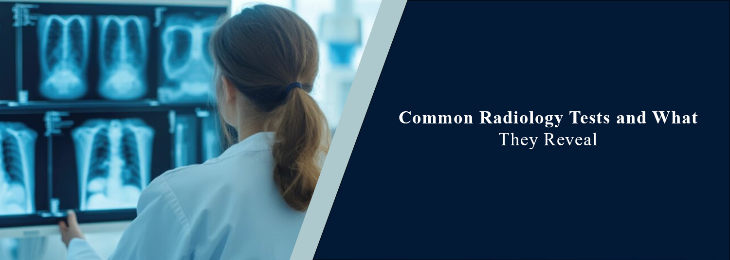
Radiological imaging methods allow
doctors to view structures within the body without invasive procedures. They
can be used to assess bones, organs, blood vessels, and soft tissue. A variety
of radiological imaging techniques can be used depending on symptoms, medical
history, and the condition being investigated. Knowing which radiology
investigations are common, and why they may be used, can be very helpful for
patients undergoing the investigations. At Molecular Diagnostics and Therapy,
diagnostic imaging allows doctors to assess a variety of conditions in patients
to make informed decisions about patient care.
What Are Radiology
Tests?
Radiology investigations allow doctors
to view structures within the body. They can be used to identify abnormalities,
monitor current conditions, and determine how well treatments are working on
certain diseases. There are many types of medical imaging investigations.
X-rays, ultrasounds, magnetic fields, or radioactive tracers may be used. Some
investigations are quick and non-invasive, whilst others may require contrast
dyes and specific preparation. Radiology investigations are generally carried
out if more information is required than can be obtained from a physical
examination or simple laboratory tests.
Common Types of
Radiology Tests and Their Uses
1. X-ray
X-rays are a common radiological
investigation. They use ionising radiation to create images within the body.
X-rays are very useful for viewing bones.
What does it reveal?
Broken bones, joint abnormalities,
infections within the chest, changes in the lungs, and dental problems can be
identified. X-rays of the chest can be used to analyse the lungs and heart.
X-rays of bones can be used to analyse injuries and changes in bones. The
investigations are typically quick. Most X-rays do not require special
preparation.
2. Ultrasound
Ultrasound scans use high-frequency
sound waves to create real-time images of organs, tissues and blood flow.
Unlike X-rays or CT scans, no ionising radiation is used.
What does it reveal?
Organ imaging can be performed within
the abdomen, pelvis, thyroid, breast, kidney and soft tissues. Cysts, certain
mass growths, fluid build-up, gallstones, and structural changes can be seen.
Doppler ultrasound can be used to analyse blood flow in blood vessels.
Ultrasound can be used in pregnancy to monitor baby development.
3. Computed
Tomography (CT) Scan
CT scans work by using X-rays and
computer programmes to create detailed cross-sectional images. More information
can be seen in a CT scan than in a simple X-ray.
What does it reveal?
CT scans can be used to analyse injury,
internal bleeding, infection, tumours, lung abnormalities and certain
abnormalities within the abdomen. CT scans can be used to image the brain,
chest, abdomen, pelvis and bones. Multiple structures can be imaged within one
scan. In some cases, contrast dyes may be used in order to clearly visualise
blood vessels, organs and certain abnormalities.
4. Magnetic Resonance
Imaging (MRI)
MRI scans use strong magnetic fields
and radio waves to create detailed images of organs and soft tissues. No
ionising radiation is used.
What does it reveal?
MRI scans are particularly useful in
imaging the brain, spinal cord, joints, muscle, ligaments and internal organs.
Conditions such as disc problems in the spine, ligament damage, abnormalities
in the brain, certain tumours and changes in soft tissue can be identified. In
some cases, contrast dyes are used in MRI scans to allow extra diagnostic
information to be gained.
5. Mammography
Mammograms are a type of specialised
imaging for the breasts. They use low-dose X-rays.
What does it reveal?
Abnormalities within the breast can be
seen. This includes mass growths, distortion, and microcalcifications.
Mammograms can be used as a screening tool for breast cancer in suitable
people. They can be used to investigate symptoms such as mass growths in the
breast and changes to the nipple. If any suspicious findings are found, further
imaging (ultrasound) or a biopsy may be recommended.
6. PET-CT Scan
PET-CT scans allow metabolic and
anatomical imaging. Small amounts of radioactive tracers are used to see how
active different tissues within the body are.
What does it reveal?
PET-CT scans are commonly used in
cancer to determine the activity of the disease, to see how far it has spread
and how well treatments are working. They may be used in specific neurological
and cardiac conditions. Different tracers can be used depending on what is
being analysed in the scan. For example, FDG PET-CT scans allow areas with high
glucose metabolism to be seen.
7. Bone Densitometry
(DEXA Scan)
Bone density can be measured using DEXA
(Dual Energy X-ray Absorptiometry) scans. Low-dose X-rays are used.
What does it reveal?
DEXA scans allow assessment of bone
strength. Osteoporosis and reduced bone density can be identified. This is very
useful in determining the risk of fractures in patients at risk. This includes
elderly patients and post-menopausal women. This allows doctors to determine
whether preventative measures or treatments may be required.
Why Are Radiology
Tests Important?
Radiological investigations allow
information to be obtained that cannot be obtained from physical examination.
They allow doctors to locate, measure, and understand abnormalities within
patients. They can be used to help make early diagnoses of disease, assess
injuries, guide treatments, and determine how well treatments are working.
Radiological investigations can also allow doctors to decide if further tests
or specialist referrals may be required. However, a definitive diagnosis may
not be able to be made based on radiological imaging. Further blood tests,
clinical examination, biopsies, or imaging may be required to confirm findings.
How Is the Right
Imaging Test Selected?
Depending on the suspected condition,
symptoms, medical history, and the area of the body being examined, different
radiological investigations may be performed. An X-ray may be performed if
there is suspected damage to bones. However, if detailed information is
required on damage to discs and ligaments in the spine, an MRI may be
performed. An ultrasound may be preferred to image certain organs in the
abdomen/pelvis. If there is complex damage, a CT scan may be recommended. Other
factors to consider when choosing a suitable investigation include pregnancy,
kidney function, implants, exposure to radiation, and the requirement of
contrast dyes. If patients are pregnant/ may be pregnant, have allergies,
kidney problems, or medical implants, they should inform the radiological team.
Conclusion
Radiological investigations are very important in diagnosis and patient care. A range of different investigations (X-ray, ultrasound, CT scans, MRI, mammograms, PET-CT scans, and DEXA scans) allows different information to be obtained. Understanding why certain diagnostic imaging investigations are performed and what information can be obtained can help patients prepare for investigations. In Molecular Diagnostics and Therapy, appropriate investigations are performed so that medical conditions can be assessed in patients and suitable care plans can be made.
Frequently Asked Questions
In News
Get Our Mobile App
for Easy Access
Book tests, view reports, and manage your health records on the go. Experience convenient healthcare with Molecular Diagnostics and Therapy.
- ✔ Book tests & home collection
- ✔ View reports instantly
- ✔ Track health history
- ✔ Get notifications & reminders
- ✔ Easy appointment management