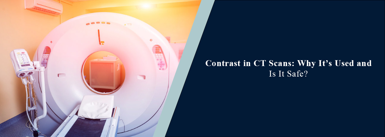
Contrast in CT Scans: Why It’s Used and Is It Safe?
Talk to Health Expert

Dr. Nikunj Jain
Co-Founder and HOD - Nuclear Medicine ,MBBS, DRM, DNB, FEBNM, FANMB, Dip. CBNC
CT scans allow doctors to view various internal structures within the
body, including organs, bones, blood vessels, and soft tissues. In some
instances, contrast media may be used to enhance the images taken in a CT scan.
Using contrast media in a CT scan means that abnormalities can be identified,
blood flow assessed, and the severity of certain diseases understood. Many
people may have questions about having contrast in their CT scan. Knowing how
the contrast works, how the scan will be performed, and what precautions should
be taken can help people feel more at ease.
At Molecular Diagnostics and Therapy, CT scans are used as part of
diagnostic investigations. Whether or not contrast media is used in your CT
scan depends on what area of the body is being investigated and what
information your doctor wants to gather.
What Is Contrast in a
CT Scan?
Contrast media is used in certain CT
scans to allow clearer views of internal structures. This allows different
tissues to be distinguished and makes it easier to identify any abnormalities.
In CT scans, iodine-based contrast
media is most commonly used. Contrast may be delivered via the intravenous (IV)
route. In some instances, contrast may be given orally. In selected instances,
contrast may be given via other routes. Unlike general medications, contrast
media in CT scans is mainly used to improve the quality of diagnostic images.
This allows radiologists to have a clearer view of organs, blood vessels, and
other structures in the body.
Why Is Contrast Used
in CT Scans?
Whilst useful information can be gained
from CT scans without contrast media, using contrast media can make it easier
to investigate certain conditions. Depending on the type of scan, contrast
media can be used for the following reasons.
1. To Examine Blood
Vessels
Contrast media allows blood vessels to
be viewed more clearly on imaging. For example, in CT angiography, contrast
media can be used to allow investigation of blood vessels and identify any
narrowing, blockage, aneurysm, or abnormal blood flow.
2. To Identify
Tumours and Abnormal Growths
Using contrast media in CT scans can
allow investigation of abnormal growths in different organs. How contrast media
is taken up and leaves the tissues can give useful information about blood
supply to tissues. This can allow investigation of tumours, identification of
abnormalities, and understanding how severe certain cancers are. However, it
may not always be possible to determine if growths are cancerous from a CT
scan.
3. To Detect
Infections and Inflammation
Contrast media may be used to identify
areas of inflammation, infection, or fluid buildup in the body. It can be
useful in investigating intra-abdominal infections, abscesses, and certain
inflammatory diseases.
4. To Examine
Internal Organs
Using contrast media in a CT scan can
allow better investigation of organs in the body (liver, kidneys, pancreas,
spleen, intestines). Structural changes, abnormal blood supply, and other
findings may be identified more easily.
5. To Evaluate
Injuries and Internal Bleeding
In certain emergencies, contrast-enhanced CT scans may allow doctors to
identify internal bleeding, organ, and blood vessel damage.
What Is the
Difference Between a CT Scan With and Without Contrast?
Contrast CT scans include contrast media, whereas non-contrast CT scans
do not. Non-contrast CT scans produce detailed images of internal structures.
They may be used to investigate certain kidney stones, bone injuries and
abnormalities in the lungs. Contrast CT scans allow certain tissues, organs or blood
vessels to be seen more clearly. Contrast CT scans may be recommended to
investigate tumours, infections, intra-abdominal conditions and abnormalities
in blood vessels. Depending on the symptoms, medical history and reason for the
scan, either a contrast or non-contrast CT scan may be most appropriate.
How Is a CT Scan With
Contrast Performed?
Contrast CT scans are generally quick
and performed by trained imaging staff. Before the scan, you may be asked about
allergies, previous reactions to contrast, kidney conditions, medications, and
other relevant medical conditions. Depending on the scan, you may be required
to have a blood test to check kidney function.
If contrast media is to be administered
intravenously, a cannula will be placed within a vein (usually in the arm).
Contrast media is then administered via the cannula, either by hand or by
injection. During administration of contrast media, you may feel a mild warming
sensation, a metallic taste in your mouth, and/or an urge to pass urine. These
feelings usually last for a short time.
Once the scan begins, you will be
placed on the CT scanning bed. As images are taken, you will be moved along the
machine. It is important to be as still as possible and follow breathing
instructions. Once the scan has been completed, if you had a cannula in place,
it may be removed. Usually, you can return to normal activities, unless
otherwise instructed by your treating doctor.
Is CT Scan Contrast
Safe?
In general, with correct
administration, iodine-based contrast media in CT scans is safe for most
people. However, as with other medical substances, some people can experience
side effects. Factors in a patient’s medical history must be considered to determine
if contrast media should be used, and whether there are any risks involved.
Common and Mild Side
Effects
Some patients may experience temporary effects such as:
- A
warm or flushed sensation during injection
- A metallic taste in the mouth
- Mild nausea
- A temporary feeling of warmth or discomfort
These effects usually resolve without treatment.
Allergic-Like
Reactions
Some people may experience an allergic
reaction to iodine-based contrast media. Symptoms can include itchiness,
rashes, hives, and swelling. Severe allergic reactions are rare but can include
breathing difficulties, large swelling and/or a drop in blood pressure. Imaging
centres should be able to recognise and manage these reactions. If you have had
an allergic reaction to iodine-based contrast media previously, please let the
imaging team know.
Kidney-Related Risks
Special considerations must be made for
people with significantly impaired kidney function or acute kidney conditions.
For some people, there is an increased risk of kidney damage in relation to
contrast media, especially people with severe kidney impairment or acute kidney
injury. Your doctor may assess your kidney function results and decide whether
contrast media is appropriate, or if an alternative investigation is required.
Who Should Take Extra
Precautions Before a Contrast CT Scan?
Although contrast-enhanced CT scans are
commonly performed, some people may require extra investigations to be
performed prior to a scan. This may apply if you have:
- A
history of an allergic-like reaction to iodinated contrast
- Chronic kidney disease or acute kidney injury
- Certain thyroid conditions, particularly when
iodine exposure may affect planned thyroid treatment
- Pregnancy or possible pregnancy
- Other medical conditions or medications that
may influence the imaging decision
If you are currently taking Metformin,
please advise your doctor and imaging team. Depending on kidney function, acute
kidney injury, and clinical guidelines, Metformin may need to be stopped
temporarily. However, you should not stop taking Metformin without medical
advice.
How to Prepare for a
CT Scan With Contrast
Depending on the type of CT scan and
type of contrast media, preparation may vary. Please ensure you provide full
medical history, previous reactions to contrast media, kidney-related
conditions, and current medications to the medical team. Some scans may require
you to fast for a set amount of time, and others may not. Please follow the
instructions given by the imaging centre.
If a kidney function test is requested,
please complete it within the time frame advised by your doctor/imaging centre.
If you have been given instructions on hydration, please follow these. If you
have fluid restrictions due to a medical condition, please follow these.
Why Choose Molecular
Diagnostics and Therapy for CT Imaging?
Choosing the right diagnostic centre is
very important. Molecular Diagnostics and Therapy offers CT scans with a
patient-focused approach. We are NABL- and NABH-accredited and have modern
diagnostic imaging equipment. Whether or not a contrast-enhanced CT scan is
performed depends on what your doctor recommends and what is required
clinically for you. Our imaging team can advise you on how to prepare, contrast
media administration, and any safety considerations.
Conclusion
Contrast media is useful in many CT scans, allowing clearer imaging of organs, tissues, and blood vessels. This can allow effective investigations of tumours, infections, injuries, and vascular conditions. Contrast media in CT scans is generally safe for most people. However, extra precautions may be needed in people with previous reactions to contrast media, kidney conditions, or specific medical conditions. Giving your full medical history and following instructions from the imaging team can help to make this a safe and comfortable procedure. Molecular Diagnostics and Therapy provides diagnostic imaging to allow informed decisions to be made by medical professionals and so that investigations can be performed in a timely manner.
Frequently Asked Questions
In News
Get Our Mobile App
for Easy Access
Book tests, view reports, and manage your health records on the go. Experience convenient healthcare with Molecular Diagnostics and Therapy.
- ✔ Book tests & home collection
- ✔ View reports instantly
- ✔ Track health history
- ✔ Get notifications & reminders
- ✔ Easy appointment management