
Can Diagnostic Imaging Help Detect Rare Diseases?
Talk to Health Expert

Dr. Nikunj Jain
Co-Founder and HOD - Nuclear Medicine ,MBBS, DRM, DNB, FEBNM, FANMB, Dip. CBNC
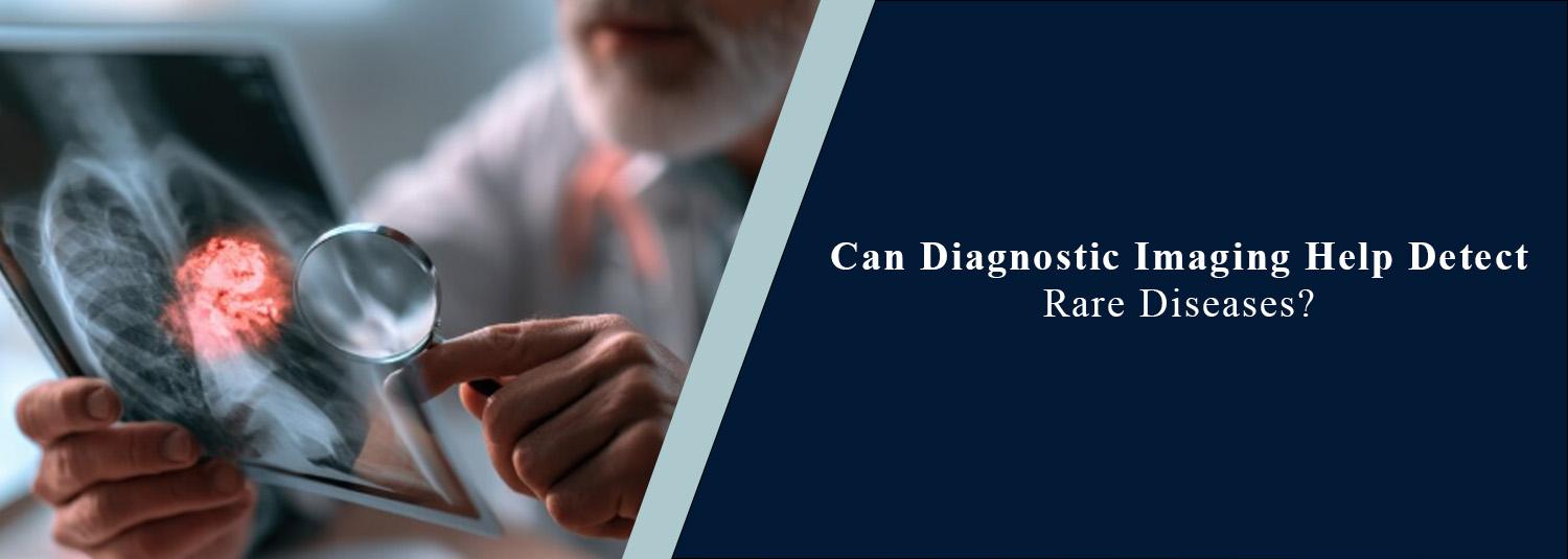
Some rare diseases have symptoms that
resemble more common health problems, making them difficult to diagnose. In
some cases, doctors need multiple tests to determine the cause of patients'
symptoms. Sometimes diagnostic imaging provides clues and reveals abnormal
changes in organs, tissues, or body functions that standard tests don't show.
When physicians require additional
information to locate abnormal tissue or to inform their understanding of
disease activity or its effects on various parts of the body, they order
advanced imaging, including an FDG PET-CT scan, a whole body PET-CT scan, and
other special scans. The use of advanced imaging and nuclear medicine services
at Molecular Diagnostics and Therapy aids physicians in the determination of a
variety of medical conditions and in planning treatment.
What Are Rare
Diseases?
Rare diseases are diseases that occur
in a fairly small number of people. There are thousands of rare diseases, and
they impact almost any part of the body. The causes of some rare diseases are
related to the genetic makeup, and for others it is related to an immune
response, metabolism disorder, a problem with the nervous system, or another
organ or tissue. The symptoms differ in each instance. Many rare diseases are
not so common and have symptoms that are confused with more common diseases,
and this complicates the diagnosis.
Why Can Rare Diseases
Be Difficult to Diagnose?
It is also one of the biggest
challenges that symptoms aren't specific to one particular disease. For
instance, in numerous situations, there is unexplained weight loss, persistent
pain or weakness, fever, swelling, or changes in organ function. Doctors then
need information provided by blood tests, genetic testing, biopsies, imaging,
and other tests to make a diagnosis. However, imaging alone is not usually
sufficient to diagnose a rare disease. Instead, once it's in, it provides
details that contribute to the physician's understanding of what is happening
in the body and determine if further testing is necessary.
How Can PET-CT Help?
PET-CT integrates details of anatomy
with information on biological activity. The patient is given an injected
amount of radioactive material, and the PET part of the machine measures the
pathological distribution of the radioactive waste. This CT information gives
details regarding the areas being studied and their structure.
Fluorodeoxyglucose (FDG) is a substance
similar to glucose that is used in an FDG PET-CT scan. Increased FDG uptake is
seen in some tissues and disease processes. Sometimes this information is
useful in helping the doctor determine where there may be abnormally active
metabolism that needs to be further studied. Increased uptake of FDG, however,
is not a confirmation that cancer is present. Other diseases and illnesses,
such as infection and inflammation, also exhibit more activity. That is why it
is important to take the PET-CT results into account along with all the other
test results and the patient's clinical history.
What Is a Whole Body
PET-CT Scan?
A whole body PET-CT scan is a single
scan that's used to screen a wide area of your body. It gives information on
several regions, depending on the clinical situation and scan protocol. In some
cases, it is helpful to get information about the distribution of abnormal
activity in patients who present with undiagnosed symptoms or those who have an
established diagnosis that includes more than one body part. In these patients,
a whole-body examination is needed. It also assists physicians to evaluate
whether the disease known to them is localized or if other areas of the body
should be further scrutinized. Whether or not a whole-body PET-CT should be
used is dependent on the clinical enquiry.
Can PET-CT Detect
Rare Cancers?
Sometimes, if a cancer is rare, it is
possible to detect it with the same imaging tests that are used for most
cancers. PET-CT gives more information about metabolism and helps find
suspicious areas. In certain cancer patients, an oncology PET-CT scan helps a
physician determine the stage of the disease, check for metastasis, and/or
determine response to treatment. But a PET-CT scan alone cannot rule out all
forms of cancer. However, some tumours do not show high uptake of FDG, and
non-cancerous conditions can also be active on a PET scan. Doctors might
recommend other imaging tests, lab tests, or a biopsy to make a diagnosis when
necessary.
How Does Imaging Help
With Rare Non-Cancerous Conditions?
PET-CT and other imaging techniques are
not restricted to cancer. Tissues develop abnormal changes due to certain rare
inflammatory, infectious, neurological, metabolic, and other diseases that are
detectable by specialized imaging studies. In fact, imaging reveals abnormal
activity, tissue texture changes, or even involvement of several organs. The
results offer further indications to doctors if patients have symptoms that do
not have a clear cause. This depends upon which condition is suspected, and the
imaging tests vary accordingly.
PET-CT Accuracy and
Rare Diseases
When talking about advanced imaging,
people want to know how accurate the PET-CT scan is. But there is no universal
accuracy percentage for each disease. The performance of PET-CT is influenced
by the type of disease being assessed, type of tracer, nature and dimensions of
abnormality, patient preparation, image quality, and other clinical factors.
Typical PET patterns are not identified in some rare diseases. Some diseases
look alike on imaging, and the small lesions are hard to spot. So PET-CT scan
findings must be correlated with other imaging findings and taken along with
other data and information.
What Other Imaging
Tests May Be Used?
PET-CT is only one part of diagnostic imaging. Doctors may also use CT,
MRI, ultrasound, X-rays, SPECT, or other specialised examinations depending on
the suspected condition. MRI can provide detailed information about soft
tissues and is particularly useful for many brain, spine, joint, and other
soft-tissue conditions. CT can provide detailed anatomical information and may
be useful for examining organs, bones, blood vessels, and other structures.
Ultrasound can provide real-time images and is commonly used for several organs
and soft tissues. In some cases, combining information from different tests
provides a clearer picture.
Why Is the Patient's
Medical History Important?
Imaging findings have to be understood in the right clinical context.
Doctors may consider the patient's symptoms, duration of illness, family
history, previous diagnoses, blood test results, previous scans, and treatment
history when interpreting imaging findings. This is particularly important when
investigating rare diseases because the imaging appearance may not always be
specific. Providing previous reports and relevant medical information can
therefore help the healthcare team understand the findings more effectively.
Does a PET-CT Scan
Confirm a Rare Disease?
Not always. A PET-CT scan can identify areas of abnormal activity and
provide information about where a disease process may be occurring. However, it
may not be able to determine the exact cause of that activity. Depending on the
findings, doctors may recommend additional tests such as MRI, CT, blood tests,
genetic testing, or tissue sampling. A biopsy may be needed in some cases to
confirm a diagnosis, particularly when cancer is suspected.
Conclusion
Rare diseases are hard to diagnose as
their symptoms manifest themselves in similar ways to more common diseases.
Diagnostic imaging gives doctors information about the structure and activity
of tissues and helps the doctor find out more when normal tests are
insufficient. In certain cases, an FDG PET-CT or whole-body PET-CT or an
oncology PET-CT may be helpful; however, a PET-CT should not replace other
investigations in the diagnosis of all types of rare conditions.
Numerous factors affect the accuracy of
PET scanning, such as the disease, the choice of tracer, the protocol used for
the scan, patient preparation, and the interpretation of the scan. The
combination of imaging and medical history, laboratory studies, and other
studies is the best approach. At Molecular Diagnostics and Therapy, advanced
imaging technologies are utilized to assist physicians in gaining insight into
a subject that aids in the diagnosis, disease assessment, and treatment
planning.
Frequently Asked Questions
In News
Get Our Mobile App
for Easy Access
Book tests, view reports, and manage your health records on the go. Experience convenient healthcare with Molecular Diagnostics and Therapy.
- ✔ Book tests & home collection
- ✔ View reports instantly
- ✔ Track health history
- ✔ Get notifications & reminders
- ✔ Easy appointment management
