
DOPA Test: Definition, Procedure & Uses
Talk to Health Expert

Dr. Nikunj Jain
Co-Founder and HOD - Nuclear Medicine ,MBBS, DRM, DNB, FEBNM, FANMB, Dip. CBNC
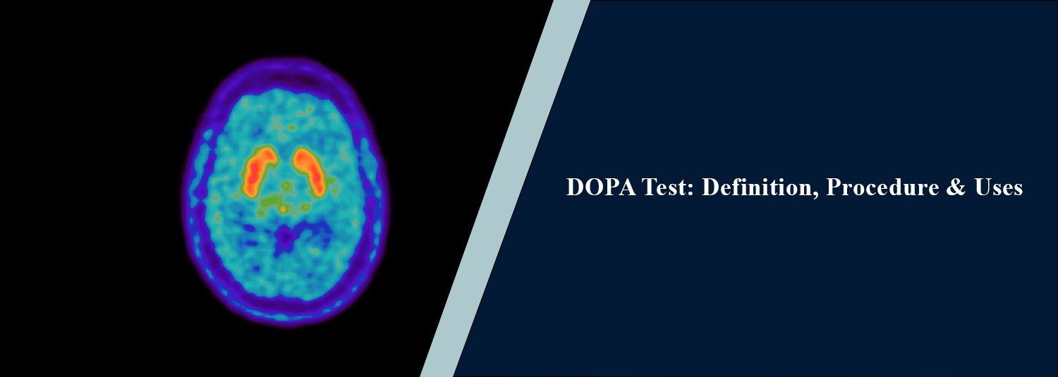
A specialised nuclear medicine imaging
test that gives information on certain biological processes in the body is
called the DOPA test, or F-DOPA PET scan / DOPA PET scan. It's based on a
radioactive compound called fluorine-18 fluorodopa (18F-FDOPA) that's
associated with dopamine metabolism. The scan can yield functional data that
won't be found through conventional imaging. Specialized nuclear medicine
imaging can, at Molecular Diagnostics and Therapy, enable physicians to use
sophisticated imaging and diagnostic methods to assess various health
conditions. Performing a DOPA PET scan will be determined by a patient's
symptoms, medical history and clinical question being evaluated.
What Is a DOPA PET
Scan?
A DOPA PET scan is a type of PET scan
that uses an 18F-FDOPA radiotracer. Once given to the patient, the tracer is
detected by the PET scanner, providing images of related biological activity.
F-DOPA relates more specifically to pathways in the brain involving dopamine.
F-18-DOPA PET could be used in a subset of neurological patients to evaluate
dopamine activity and help evaluate Parkinsonian disorders. Depending on the
disease and the purpose of the scan, applications of DOPA PET may be for the
evaluation of selected neuroendocrine and brain tumors.
Why Is a DOPA Test
Done?
There are several primary uses for DOPA
PET scans, depending on the region being studied and the suspected condition.
It can also be used in neurological imaging for the evaluation of the pathways
involved in the production of dopamine and may assist in differentiating some
other conditions with similar symptoms from the Parkinsonian syndromes.
F-DOPA PET can give information about
the biological activity and specific information about certain tumours and be
used in selected cancer-related situations. It can be especially helpful when
conventional imaging doesn't provide enough information to answer the clinical
question. This test is not a routine screening test for all neurological or
cancer problems. A specialist decides if it is appropriate for their individual
situation.
DOPA PET Scan
Procedure
Typically, the DOPA PET scan procedure
starts with an infusion of the radioisotope 18F-FDOPA, which is usually an
injection into a vein (intravenous infusion). Once injected, the tracer is allowed
to pass so it can reach and be concentrated in the tissue under evaluation. The
patient is then placed on the PET scanner, and the radioactivity is detected to
provide a detailed image. When pictures are taken, the patient must not move to
make sure the image is clear. In some examinations, the patient might be asked
to prepare for the scan beforehand, while the centre may also provide
instructions to follow during the imaging process itself. These images are then
analysed and interpreted by a skilled and trained nuclear medicine doctor. PET
imaging can also offer functional data, and when clinically indicated, PET/CT
can combine functional and anatomical information from CT.
DOPA PET Scan for
Parkinsonian Disorders
An important use of F-DOPA PET scan is
to measure how active the dopamine system is in the brain. Parkinsonian
disorders may sometimes be hard to differentiate from other diseases that may
also produce tremor, stiffness, or movement-related abnormalities. With the
reduced dopamine activity shown by F-18-DOPA PET, the scan could also
potentially help in the clinical assessment of Parkinson's disease and other
Parkinsonian-related disorders. However, the scan alone cannot provide a
diagnosis; its images must be interpreted by a trained nuclear medicine
physician in the context of the patient's symptoms and other findings. To
establish a diagnosis, doctors will consider the person's symptoms and look for
signs in their neurological assessment, medical history, and other tests.
DOPA PET Scan in Cancer
Evaluation
In selected tumour-related
investigations, F-DOPA PET can be used as well. Amino-acid PET tracers (e.g.
FDOPA) may be used to assess the type of certain brain tumours, including
gliomas. It can be used to give information on the biology of the tumour and
may be used to supplement MRI scans and other imaging techniques. The value of
DOPA PET differs with the tumor type and with the clinical question. This
should only be done if the treating professional or nuclear medicine doctor
recommends it.
How Should You
Prepare for a DOPA Test?
The preparation will depend on the
reason for the scan and the “protocol” of the centre. Important history to
capture consists of the patient's current medications, medical or surgical
history, prior imaging studies, and any treatment received. The nuclear
medicine staff will give you special instructions on diet, fluids, medication,
and other preparation instructions. These instructions should be followed
carefully and not changed without guidance from the healthcare provider or
nuclear medicine team.
Is the DOPA PET Scan
Safe?
Exposure in DOPA PET comes from a small
amount of radiation from the radioactive tracer. When carefully prescribed and
administered, nuclear medicine procedures are generally safe, although any
risks and benefits should be weighed for the individual patient. Pregnant women
or those suspected of being pregnant or who are breastfeeding should tell the
medical team so that extra precautions are taken.
What Happens After
the Scan?
Patients will be able to return to
normal activity upon completion of the exam unless otherwise directed. The
radioactivity of the radiotracer slowly fades over time, and the radiotracer is
excreted from the body. With certain nuclear medicine procedures, patients may
be required to consume fluids, depending on their medical status and the
procedure.
Why Choose Molecular
Diagnostics and Therapy?
Molecular Diagnostics and Therapy
offers cutting-edge diagnostic and nuclear medicine services to assist in the
interpretation of various health conditions. Specialist molecular imaging can
supply functional information in addition to normal anatomical imaging.
Depending on the patient's condition and the question to be answered, a DOPA
PET scan may or may not be considered. A specialist will be able to suggest
what imaging techniques will be necessary and what the results will mean,
taking into account the general condition of the patient.
Conclusion
DOPA test (also known as F-DOPA PET scan) is a special kind of nuclear medicine study that can get information about dopamine activity and other biochemical processes. It is used in some neurological examinations (e.g., Parkinsonian disorders) and in some investigations related to tumors. The DOPA PET scan procedure generally involves injecting the 18F-FDOPA radiotracer, followed by PET imaging to evaluate the relevant biological activity. The test is a special test and should be used if it is indicated for a certain clinical situation. Molecular Diagnostics and Therapy provides advanced diagnostic and nuclear medicine services to support appropriate evaluation and care for patients.
Frequently Asked Questions
In News
Get Our Mobile App
for Easy Access
Book tests, view reports, and manage your health records on the go. Experience convenient healthcare with Molecular Diagnostics and Therapy.
- ✔ Book tests & home collection
- ✔ View reports instantly
- ✔ Track health history
- ✔ Get notifications & reminders
- ✔ Easy appointment management
