
What Is a Brain MRI and When Do You Need One?
Talk to Health Expert

Dr. Nikunj Jain
Co-Founder and HOD - Nuclear Medicine ,MBBS, DRM, DNB, FEBNM, FANMB, Dip. CBNC
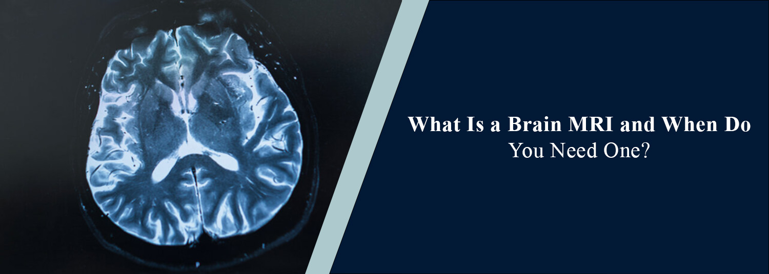
A Brain MRI can be used to examine the
brain in detail. If you have persistent symptoms, such as headaches, dizziness,
seizures, visual changes, or any other neurological symptoms, your doctor may
recommend a brain MRI, which is one of the most detailed types of imaging.
Doctors can use a brain MRI to look at
the brain and detect any abnormalities within the brain tissues, blood vessels,
and surrounding areas. Unlike a CT scan, this imaging technique uses magnetic
fields and radio waves, and therefore does not expose the patient to ionizing
radiation. At Molecular Diagnostics and Therapy, brain MRIs are used to help
doctors make informed decisions about neurological conditions.
What Is a Brain MRI?
A Brain MRI is a non-invasive imaging
test that uses strong magnetic fields and radio waves to create detailed images
of the brain. This allows doctors to look at grey matter, white matter,
cavities, and certain blood vessels within the brain. As soft tissues are
visible in detail, this technique can be very useful in diagnosing conditions
that may not be clearly seen using other imaging techniques. Depending on your
symptoms and suspected condition, a contrast agent may or may not be used. If
contrast is used in a brain MRI, it is typically a gadolinium-based solution
that is injected into a vein. This can allow certain structures and
abnormalities to be more visible within the brain.
What Are the Common
Uses of a Brain MRI?
A brain MRI may be indicated in a
variety of neurological conditions. The reason for the MRI depends on the
symptoms and medical history of the patient.
1. Evaluating
Persistent or Unusual Headaches
While most headaches do not indicate
serious brain conditions, if you are experiencing persistent, unusual,
worsening headaches, or neurological symptoms, a brain MRI may be indicated.
2. Detecting Brain
Tumours
A brain MRI can give detailed images of
tissues within the brain. It can help identify abnormal growths in the brain.
If a brain tumour is suspected, it can help determine the size, location, and
extent of involvement of surrounding areas. If contrast is used, extra
information may be provided about certain types of tumours. However, it may not
be possible to determine the exact type of tumour from imaging alone.
3. Investigating
Stroke
A brain MRI can help to identify areas
of damage to brain tissues due to reduced blood flow or bleeding. Certain types
of brain MRI, for example, diffusion-weighted imaging, are very useful in
identifying early changes in patients with an ischaemic stroke. If a patient is
in an urgent situation, a CT scan may be performed first. This is a very quick
test widely used to identify possible acute bleeding in the brain.
4. Diagnosing
Seizures and Epilepsy
A brain MRI may be used to identify
structural changes in the brain that may cause seizures. It may identify
certain developmental changes, scarring, or lesions. It is often performed
alongside clinical information and other investigations, such as an EEG.
5. Evaluating
Multiple Sclerosis
MS is an autoimmune disease, meaning
that the immune system attacks the protective sheaths surrounding nerves in the
brain and spinal cord. Using MRI, areas of inflammation and damage (lesions)
can be identified. This can help with diagnosing multiple sclerosis and
monitoring the progression of the disease.
6. Investigating
Memory and Cognitive Changes
If a patient is having memory issues,
confusion, or unusual cognitive changes, a brain MRI may be indicated. A brain
MRI can help identify structural changes, previous strokes, or volume loss in
the brain. However, it is important to note that a brain MRI alone cannot
determine the exact cause of memory loss/dementia in all patients. Other
investigations and a clinical assessment may be required.
7. Examining Brain
Blood Vessels
Using specific imaging techniques, such
as MRA (Magnetic Resonance Angiography), a brain MRI can be used to look at the
blood vessels in and around the brain. This may be used to investigate certain
conditions, including aneurysms, narrowing of blood vessels, and vascular
abnormalities.
When Do You Need a
Brain MRI?
Not all patients with headaches,
dizziness, or neurological symptoms require a brain MRI. If symptoms, physical
examination, or medical history indicate that a detailed brain image would be
helpful, a brain MRI may be indicated. In some cases, a brain MRI may be
indicated if:
- Persistent
or progressively worsening headaches
- Repeated seizures or unexplained episodes of
loss of awareness
- Unexplained weakness or numbness
- Changes in vision, speech, or coordination
- Memory loss or unusual cognitive changes
- Suspected brain tumours or structural
abnormalities
- Follow-up of certain previously diagnosed
neurological conditions
If you have sudden weakness in the
face, on one side of your body, speech difficulties, sudden visual loss, or
severe unexpected headaches, this may be an emergency. Seek emergency medical
help.
How Is a Brain MRI Procedure
Performed?
A brain MRI is generally painless.
Unless a contrast agent is required, no injections are given.
Before the Scan
Before the scan, you are asked questions by the imaging team about your
medical history, previous operations, and any metal implants or devices. If you
have a pacemaker, cochlear implant, aneurysm clip, or any other implants, you
should inform the imaging team. You are asked to remove all jewellery, watches,
and other metal items. If a contrast agent is to be used, the doctor may want
to check kidney function and whether you have had any reactions to contrast
agents in the past.
During the Scan
You lie on a movable bed that enters
the MRI machine. A special head coil may be placed around your head to produce
clear images. During the scan, you may hear loud knocking or clicking noises.
You may be given earplugs or headphones to help reduce the noise. It is
important to remain very still during the scan; otherwise, images may not be as
clear. In certain sequences, the imaging team may ask you to follow simple
instructions.
After the Scan
Once the required images have been
taken, you can usually resume normal activities. If contrast was used, the
imaging team may give you further instructions depending on your medical
condition. A radiologist analyses the images and produces a report for the
referring doctor. The referring doctor explains the findings to you and makes
recommendations if necessary.
How Long Does a Brain
MRI Take?
The length of the scan may vary
depending on the type of imaging that is required. A standard brain MRI may
take between 20 and 45 minutes. If contrast is used, or specific imaging is
required, it may take longer. To allow clear images to be produced, try to stay
as relaxed and still as possible during the scan.
Is a Brain MRI Safe?
A brain MRI is generally a safe
procedure, as long as the correct precautions are taken. As no ionising
radiation is used, it is different from X-rays and CT scans. However, as strong
magnetic fields are used, interactions with metal objects or medical devices
may occur. It is very important to be accurate with your information, and
inform the imaging team if you have implants, previous operations, or may have
any metal pieces in your body. Some individuals may find the claustrophobic
environment uncomfortable. If you suffer from claustrophobia, make sure to
inform the imaging team in advance so suitable arrangements can be made. If a
contrast agent is used, those with severe kidney issues or a history of
reactions to contrast agents may require further assessment.
What Is the Cost of a
Brain MRI Scan?
The cost of a brain MRI depends on the
type of scan, whether contrast is used, the type of imaging, and the diagnostic
centre. A standard scan may be different in cost compared to a
contrast-enhanced scan or a scan with angiography. Before making an appointment,
confirm with the diagnostic centre what type of scan is performed, whether
contrast is used, and any preparation required.
How to Prepare for a
Brain MRI
Depending on what type of brain MRI is
to be performed, preparation may vary. Inform the imaging team of any implants,
previous operations, allergies, kidney issues, and the medications you are
currently taking. Not all standard brain MRIs require fasting. However, if you
are having a contrast-enhanced scan, or a scan with sedation, there may be specific
instructions. Always follow the instructions given by the imaging centre. If
you get anxious in small, enclosed spaces, make sure to inform them in advance.
Brain MRI Services at
Molecular Diagnostics and Therapy
Imaging can be very useful in assessing
neurological conditions. At Molecular Diagnostics and Therapy, MRI scans are
performed by our team to help doctors examine the brain and other parts of the
body. We are NABL and NABH accredited and are equipped with modern diagnostic
imaging equipment. Once the results of a brain MRI are obtained, they are
reviewed by qualified radiologists. The results are then given to the referring
doctor for assessment. The type of MRI is chosen depending on the symptoms,
medical history, and recommendations from your doctor.
Conclusion
A brain MRI is a very useful diagnostic test. It can be used to investigate conditions such as brain tumours, stroke, seizures, multiple sclerosis, and certain causes of persistent neurological issues. It does not expose patients to ionising radiation. However, not all patients with headaches or dizziness need a brain MRI. The decision should be made based on clinical assessment and what information is required by your doctor. At Molecular Diagnostics and Therapy, we offer diagnostic imaging to help assess neurological conditions, so that patients can make informed decisions to improve their healthcare.
Frequently Asked Questions
In News
Get Our Mobile App
for Easy Access
Book tests, view reports, and manage your health records on the go. Experience convenient healthcare with Molecular Diagnostics and Therapy.
- ✔ Book tests & home collection
- ✔ View reports instantly
- ✔ Track health history
- ✔ Get notifications & reminders
- ✔ Easy appointment management