
PSMA Test: Types, Uses, and Procedure
Talk to Health Expert

Dr. Nikunj Jain
Co-Founder and HOD - Nuclear Medicine ,MBBS, DRM, DNB, FEBNM, FANMB, Dip. CBNC
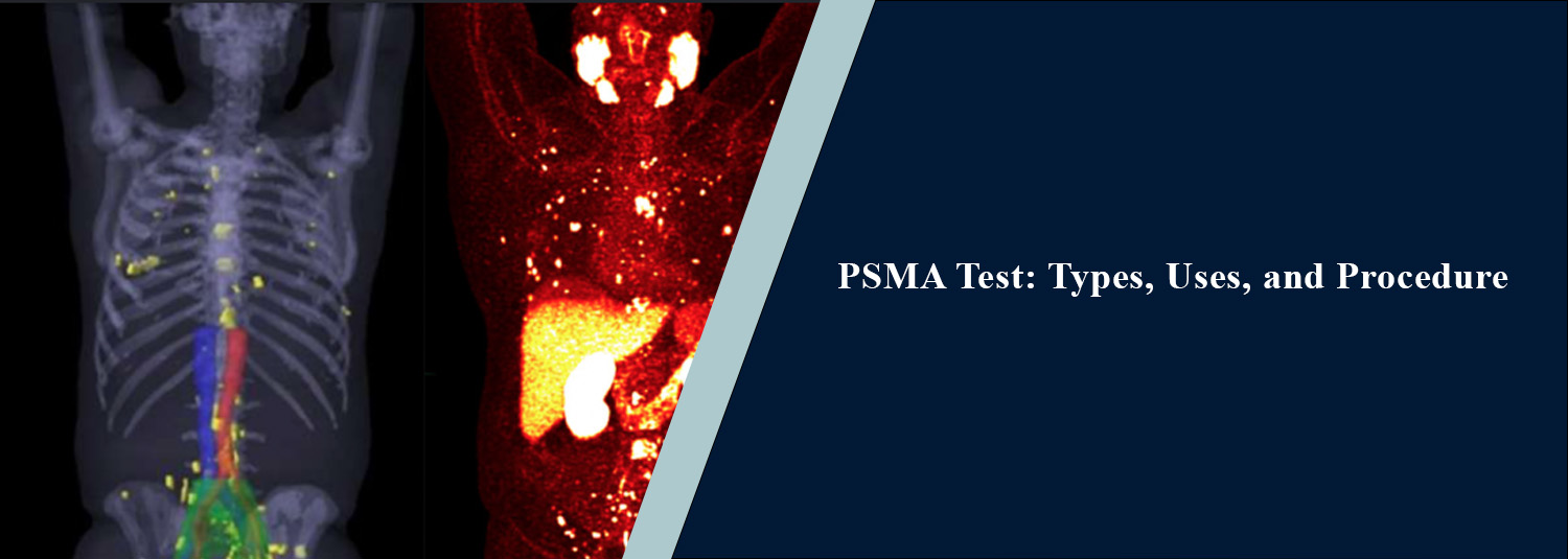
A PSMA Test is an advanced nuclear
medicine diagnostic technique that has proven useful in assessing prostate
cancer. A PSMA PET scan uses trace amounts of a radioactive tracer compound to
target prostate-specific membrane antigen (PSMA), which is up-regulated on many
prostate cancer cells. In combination with CT, the scan can help doctors
determine the location and extent of prostate cancer tumours. At Molecular Diagnostics and Therapy, advanced nuclear medicine and PET-CT diagnostics help
assess and monitor prostate cancer, as directed by your treating doctor.
What Is a PSMA Test?
PSMA stands for Prostate-Specific
Membrane Antigen. It is a protein on the surface of prostate cancer cells and
can be up-regulated significantly in comparison to normal prostate cells. A
PSMA PET scan involves injecting a PSMA-targeting radiotracer through an
intravenous (IV) line. The radiotracer then binds to cells that carry PSMA,
enabling the PET scan to detect where the cancerous cells are.
When used in combination with a CT
scan, the nuclear medicine PET (Positron Emission Tomography) image identifies
regions where the radiotracer has accumulated, while the CT (Computed
Tomography) image enables the radiologists to identify the specific anatomical
location and surrounding structures. Together, both scans provide complementary
information and give doctors a better understanding of where suspected
cancerous tumours might be.
Types of PSMA PET
Scans
A variety of PSMA radiotracers may be
used depending on availability, clinical need, and other factors. Gallium-68
PSMA and Fluorine-18 PSMA are examples of radiotracers that are occasionally
used. While these radiotracers may have differences in radioactive isotope
(radioactive element), they are generally PSMA-targeted and are selected by the
nuclear medicine team based on the clinical indication and available equipment.
What Is a PSMA PET-CT
Scan Used For?
A PSMA PET-CT scan may be recommended in different situations involving
prostate cancer.
Initial Staging
In some men presenting with newly
diagnosed prostate cancer, a PSMA PET-CT scan may help determine whether
prostate cancer has spread beyond the prostate gland. This is an important
consideration when determining the staging of prostate cancer and, therefore,
which treatment options are appropriate.
Detecting Recurrent
Prostate Cancer
PSMA PET-CT scanning may also be used
to help detect prostate cancer that recurs (returns or progresses) after
initial treatment for prostate cancer, particularly when the PSA
(Prostate-Specific Antigen) level rises after surgery or radiotherapy for
prostate cancer. In such cases, the scan may help identify regions of recurrent
prostate cancer that would not typically be apparent with conventional imaging.
Assessing Spread of
Disease
PSMA PET-CT scanning can provide
information about the spread (metastases) of prostate cancer, including
involvement of lymph nodes (common sites of prostate cancer metastasis) and
bones (another common site of prostate cancer metastasis). Knowing whether
cancer has spread may also help determine the relative appropriateness of
different treatment options.
Before PSMA-Targeted
Therapy
PSMA imaging may also be used to help
determine whether a man is considered appropriate for PSMA-targeted radioligand
therapy. The PSMA PET scan helps determine whether the cancerous lesions
express enough PSMA for the radioligand to bind effectively for the proposed
treatment.
PSMA PET Scan
Procedure
The procedure is generally simple but
has several steps. A patient is first asked to present relevant medical
documents and information about their prostate cancer diagnosis, any prior
treatments for prostate cancer, PSA (prostate-specific antigen) results, and
any prior imaging. A cannula is then placed by a healthcare professional, and a
small amount of the selected PSMA radiotracer is injected.
The patient is then instructed to wait
for a certain period while the tracer accumulates in the body's tissues. For
many PSMA PET scans, this waiting period is about an hour long but may be
extended depending on the facility and the radiotracer used. The patient is
then positioned on the scanning table, which moves through a PET-CT scanner to
acquire PET and CT images. This scanning procedure generally takes about 30
minutes, although because of the initial waiting period and preparation, this
is only a small part of the overall examination.
How Should You
Prepare for a PSMA PET Scan?
The preparation steps for a PSMA PET
scan may vary depending on the radiotracer and facility. In general, for most
PSMA PET-CT scans, fasting is not required, and patients are encouraged to take
their regular medication as per their physician's instructions, unless
otherwise informed. Patients are encouraged to stay well-hydrated and may even
be instructed to empty their urinary bladder at certain points during the
procedure, as some PSMA PET radiotracers are excreted in the urine. Always
follow the specific instructions provided by the nuclear medicine centre
performing the PSMA PET scan.
Is a PSMA PET Scan
Safe?
A PSMA PET-CT scan makes use of trace
amounts of a radiotracer, which is selected and administered according to
nuclear medicine protocols. Like most other imaging procedures, a PSMA PET-CT
scan is not foolproof. Regions of tracer uptake are not always cancerous, and
very small cancerous lesions may occasionally be missed. Moreover, healthy
organs typically have some tracer uptake, and so the findings must always be
interpreted in conjunction with one's PSA level, biopsies (if any), treatment
history (if any), and other relevant health information.
What Happens After
the Scan?
The scanned images are then reviewed
and reported upon by a nuclear medicine physician or imaging specialist. The
report will generally highlight any regions of increased tracer uptake. The
patient's doctor will then review the report as part of their wider clinical
assessment of the patient. Where the scan results are inconclusive, additional
imaging, lab tests, or biopsies (if applicable) may sometimes be recommended.
Why Choose Molecular
Diagnostics and Therapy?
Molecular Diagnostics and Therapy
provides advanced nuclear medicine and PET-CT diagnostics for patients who
require expert imaging. PSMA PET-CT scanning provides invaluable information
about prostate cancer localisation and spread where it is appropriate. The most
appropriate type of imaging depends on one's diagnosis, PSA level, treatment
history, symptoms, and overall clinical presentation.
Conclusion
A PSMA PET scan is an advanced nuclear medicine imaging procedure that targets PSMA to provide invaluable information about prostate cancer lesions. A PSMA PET-CT scan combines nuclear medicine and precise anatomical imaging to help determine the localisation and extent of prostate cancer tumours. This scan may be used for initial staging, detecting prostate cancer recurrence, determining the spread of prostate cancer, and determining the suitability for PSMA-targeted treatment. However, a PSMA PET-CT scan is not interpreted in isolation. The scan results must always be interpreted in conjunction with one's overall medical presentation. Molecular Diagnostics and Therapy provides advanced diagnostic imaging for the assessment and management of prostate cancer.
Frequently Asked Questions
In News
Get Our Mobile App
for Easy Access
Book tests, view reports, and manage your health records on the go. Experience convenient healthcare with Molecular Diagnostics and Therapy.
- ✔ Book tests & home collection
- ✔ View reports instantly
- ✔ Track health history
- ✔ Get notifications & reminders
- ✔ Easy appointment management
