
Nuclear Gamma Scan: Purpose, Procedure and Types
Talk to Health Expert

Dr. Nikunj Jain
Co-Founder and HOD - Nuclear Medicine ,MBBS, DRM, DNB, FEBNM, FANMB, Dip. CBNC
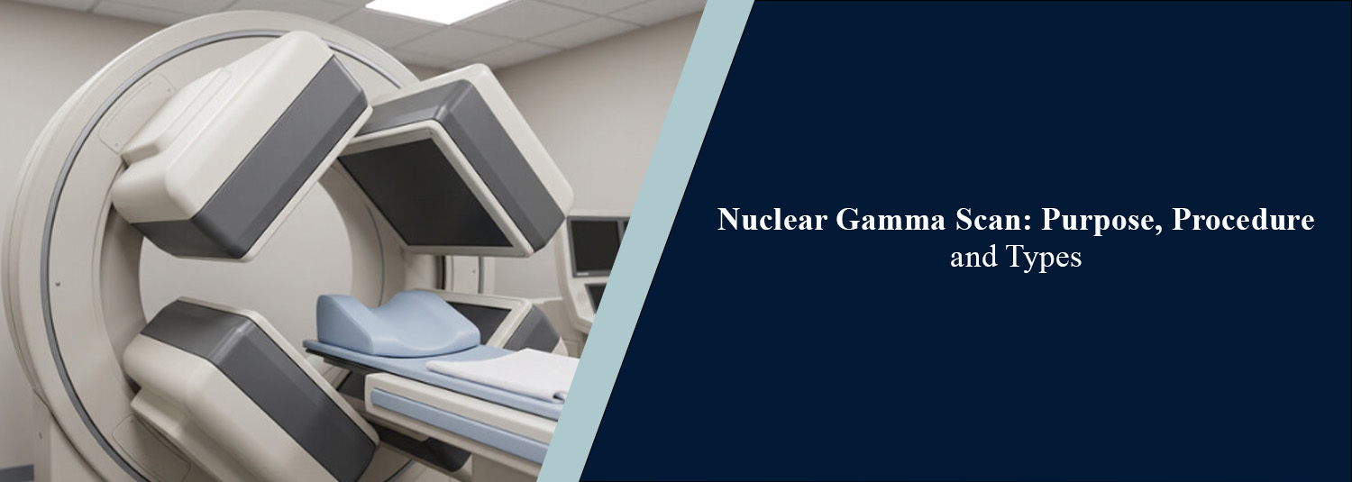
A gamma scan is an imaging technique
using nuclear medicine to assess the functioning of certain organs and tissues.
This process uses a minimal amount of radioisotopes or radiotracers in
conjunction with the camera to visualize body activities. While there are some
imaging techniques that mainly focus on showing the anatomical structures of
the body, a gamma scan can also offer indications of the functions of the
organs involved or the movement of the radiotracers through the body.
Nuclear medicine imaging is used to
help evaluate various medical conditions in Molecular Diagnostics and Therapy.
The type of scan proposed depends on the symptoms, medical history, and the
clinical question.
What Is a Nuclear
Gamma Scan?
A nuclear gamma scan, or gamma camera scan,
is a diagnostic test that identifies gamma rays that are emitted from a
radioactive substance within the body. Typically, the radiotracer is
administered via injection, although the capsule or liquid form is sometimes
used. The tracer is targeted to specific organs or tissues. Once the signal is
captured by the gamma camera, it's processed by the computer to generate
images. These images can be used to determine the functions of different organs
and detect regions of the body that require additional investigation.
Interpretation of results is considered with symptoms, examination, and other
tests.
What Are the Uses of
a Gamma Scan?
Gamma scans are used to investigate different health conditions
depending on the organ being examined. Common applications include:
- Bone
evaluation:
Helps assess certain bone abnormalities, infections, injuries, and
possible spread of cancer to bones.
- Heart assessment: Some nuclear medicine scans evaluate blood
flow to the heart muscle.
- Kidney function: Renal scans can assess kidney function,
drainage, and certain blockages.
- Thyroid evaluation: Thyroid scans help examine the gland's
structure and activity.
- Liver and gallbladder assessment: Selected scans evaluate bile flow and
gallbladder function.
- Cancer evaluation: Certain nuclear medicine studies help assess
disease activity and monitor treatment response.
The specific test is selected according to the condition being
investigated. A gamma scan does not diagnose every disease on its own.
Types of Nuclear
Gamma Scans
The term gamma scan covers several examinations. The main types include:
1. Planar Gamma Scan
This type of scan produces a
two-dimensional picture of the organ or region of interest being looked at.
Images are usually taken from fixed positions, such as front and back views.
2. SPECT Scan
Single photon emission computed
tomography (SPECT): a gamma camera is able to rotate around the patient and
produce 3D images. It can give information on where the tracer is distributed
in an organ and its distribution state at a more detailed level.
3. SPECT/CT Scan
SPECT/CT is a nuclear medicine scan
performed in conjunction with computed tomography (CT). On a SPECT scan, the
information is about the activity of the tracer, and on a CT scan, it is about
the structure. The fusion of pictures can assist doctors in pinpointing the
exact location of an irregularity more clearly.
How Is a Gamma Scan
Performed?
The procedure is different depending on
the type of scan and the organ being scanned. The general steps are as follows.
First, the healthcare team inspects the patient's medication, preparation, and
medical history. A small amount of the radiotracer is typically injected into a
vein. Some scans require the tracer to be swallowed.
Depending on the area being examined,
the patient may have to wait until the tracer reaches the area. Waiting times
vary with the type of test and can range from relatively short to hours. The
gamma camera detects the gamma rays emitted by the tracer while the patient
lies on the imaging table. Appropriate images are required, and the patient may
be asked to move or turn to get the best views. An expert then looks at the
pictures and writes a report for the referring doctor.
How Should You
Prepare for a Gamma Scan?
Prep varies with the scan obtained.
Many scans need no fasting, but some, such as hepatobiliary or cardiac stress
studies, do. Follow the instructions for your specific scan. Before the test,
tell the doctor or technologist what medications and dietary supplements you
are taking, if you have had any medical tests or surgery recently, and any
medical conditions or allergies that might affect the test.
It is important to inform the team if
you are pregnant, or may be pregnant, or are breastfeeding, as this may impact
on which and when the examination is performed. Never stop or change any
medication without your healthcare professional’s direction.
Is a Gamma Scan Safe?
The amount of radioactive substance
used in a nuclear medicine scan is carefully checked. Most diagnostic gamma
scans are non-invasive, with few side effects, and are generally well
tolerated. The amount of radiation the patient receives from the tracer is
considered when deciding whether the test is appropriate.
The benefits and risks are dependent on
the exam and/or patient situation. Any special precautions will be explained by
the healthcare staff, including those pertaining to pregnancy or breastfeeding.
Most patients can resume normal activities after the scan unless told
otherwise.
What Is the
Difference Between a Gamma Scan and a CT Scan?
A gamma scan detects signals from the
radiotracer inside the body and may be used to give information about organ
function. CT scans are imaging tests that involve using X-rays to create
detailed images of the structure of that part of the body. Some exams involve
the use of both techniques (SPECT/CT), which enables physicians to compare
information related to function with that related to anatomy. The choice will
vary depending on the medical question being studied.
Conclusion
The nuclear gamma scan can be used as
an effective diagnostic test to assess the function of certain organs and
tissues. There are different types, all offering a different amount of detail
for specific clinical needs: planar imaging, SPECT, and SPECT/CT. The
preparation time, time for the scan, and safety protocols depend upon the
examination, and so patients should follow the instructions of their healthcare
team.
Molecular Diagnostics and Therapy offers diagnostic services to support the evaluation and monitoring of health conditions. Choosing the correct test always depends on the patient's requirements and the guidance of the prescribing doctor.
Frequently Asked Questions
In News
Get Our Mobile App
for Easy Access
Book tests, view reports, and manage your health records on the go. Experience convenient healthcare with Molecular Diagnostics and Therapy.
- ✔ Book tests & home collection
- ✔ View reports instantly
- ✔ Track health history
- ✔ Get notifications & reminders
- ✔ Easy appointment management
