
Color Doppler Ultrasound: What Does It Evaluate?
Talk to Health Expert

Dr. Nikunj Jain
Co-Founder and HOD - Nuclear Medicine ,MBBS, DRM, DNB, FEBNM, FANMB, Dip. CBNC
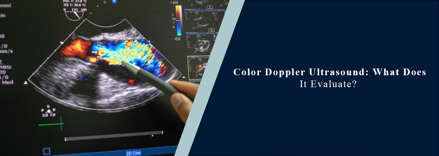
Color Doppler Ultrasound can assist in the assessment of both the structure of the body organs and the flow of blood in the arteries and veins. Color Doppler Ultrasound is a type of ultrasound, and while some ultrasound scans are used to create images of the organs and tissues, Color Doppler Ultrasound adds color to indicate the direction and speed of blood flow. This consequently makes it an important aid in the diagnosis of various vascular and organ conditions.
It's a routine test that a doctor sometimes recommends if they see signs or symptoms of swelling, leg pain, bad circulation, suspected blood clots, leg swelling, or pregnancy issues. When looking for an ultrasound scan near me, it is important to know what this form of examination measures to help you to know what to expect. The Color Doppler ultrasound is an advanced technology that utilizes experienced radiologists to ensure accurate exams and that ultimately aids in better patient care and early diagnosis at Molecular Diagnostics and Therapy.
What Is a Color Doppler Ultrasound?
A Color Doppler Ultrasound is a sophisticated ultrasound examination that also measures blood flow and uses high-frequency sound waves to produce images of the internal organs. Moving blood appears in various colors in the scan, which enables a doctor to determine whether the blood flow is normal, if there is a blockage, narrowing, or abnormal blood flow. The advantage of ultrasound is that it's non-invasive, meaning there's no radiation involved, unlike CT scans and X-rays, so it's safe to use for babies, children, or pregnant women.
Why Is a Color Doppler Ultrasound Performed?
This test is recommended to help in measuring blood circulation and diagnosing arterial and venous disease. It's frequently used to learn about lower leg swelling, pain, numbness, varicose veins, blood clots, decreased blood supply, and abdominal blood vessel disease. It is also used in a medical setting to check blood flow before and after some surgeries.
What Conditions Can It Evaluate?
Color Doppler Ultrasound can help make a diagnosis for a variety of medical conditions. It can find blocked arteries, narrow blood vessels, varicose veins, the lack of blood flow, and abnormal blood flow, as well as deep vein thrombosis (DVT) and aneurysms. It can also be used to check if blood flow is adequate to the kidneys, liver, and thyroid. If there are any circulation concerns, doctors can start treatment as soon as possible before more complications result.
Color Doppler During Pregnancy
Color Doppler Ultrasound makes a significant contribution to high-risk pregnancies. It checks blood flow from the placenta to the baby, so it is able to determine whether the baby is getting sufficient oxygen and nourishment. This test is suggested when a fetus doesn't seem to be growing at the expected rate, or when there is a pregnancy problem. The exam is painless and harmless as long as it is conducted by a trained individual.
Evaluating Heart and Blood Vessels
Colour Doppler is often used to evaluate blood flow through large veins and arteries. It aids a doctor in detecting narrowing of blood vessels, insufficient blood flow, irregularities of the valves, and circulatory diseases, which can enhance stroke risk or put somebody at risk for different cardiovascular issues.
Assessing Kidney and Liver Blood Flow
The kidneys and liver have a closer connection with blood circulation. A Color Doppler Ultrasound can assess blood flow to these organs and can detect vascular irregularities that can lead to organ dysfunction. Other tests and imaging procedures may also be used in conjunction with this information to make a complete evaluation.
Diagnosing Varicose Veins
Color Doppler Ultrasound may be performed in patients who have bulging, kinked, or painful veins. During the exam, it can tell if the valves within the veins are working correctly and if blood is moving in the proper direction. This is useful information for planning the treatment for varicose veins.
Ultrasound Scan Test Details
Patients can enquire about the details of the ultrasound scan before their visit. The method is quick and non-invasive and not painful. A gel is rubbed on the skin, and a hand-held apparatus known as a transducer is drawn across the area to be studied, or examined. The sound waves travel into the body, and back-reflected echoes are converted to detailed images by the transducer. The examination typically lasts 20-45 minutes, depending on the body part being evaluated.
Ultrasound Scan Procedure
The ultrasound scan procedure is easy and comfortable to do. Patients are typically positioned on an exam table, and the scan is performed by the radiologist or sonographer. In some instances, it may be necessary to alter your body position or have to hold your breath for a short time in order to get a clearer picture. Upon completion of the examination, the gel is removed, and patients are able to typically resume their regular daily routine right after the examination.
Ultrasound Scan Preparation
Preparation of the ultrasound scan is determined by the type of ultrasound examination. If an abdomen scan is indicated, a few hours of fasting may be necessary. Patients may be required to drink water and not urinate before a pelvic or urinary bladder scan. Precise handling of the preparation helps to enhance image quality and diagnosis.
Ultrasound Scan Benefits
Because there are numerous ultrasound scan benefits, the test is routinely performed in modern medicine. Painless, radiation-free, provides real-time imaging, evaluates blood flow, and can be repeated for follow-up examinations. It's also not harmful to kids and pregnant women. Compact ultrasound scanning is frequently one of the primary imaging tests suggested by the doctor because it is speedy, gentle, and non-intrusive.
Understanding Your Ultrasound Scan Report
Once the exams are complete, the radiologist looks through all the images and creates a report of the ultrasound. The report contains information on whether the blood flow and the organs examined are normal or not. Your doctor will discuss what he found and if any tests or treatment will be needed. It is not advisable to read into the report without medical advice.
Choosing the Right Diagnostic Centre
If you are searching for an ultrasound scan near me, choose a diagnostic centre that offers experienced radiologists, modern ultrasound equipment, accurate reporting, and quality patient care. Reliable imaging plays an important role in helping doctors make correct diagnoses and treatment decisions.
Conclusion Color Doppler Ultrasound is a valuable imaging test able to assess organ structure as well as blood circulation. It aids in the diagnosis of vascular disorders, proper monitoring of pregnancy, assistance in assessing the blood supply of the organs, and use to detect circulation disorders in advance. By understanding how the ultrasound scan works, what needs to be done in preparation for this scan, what there is to gain from going through it, and how the ultrasound scan report is read, the patient may feel secure before their exam. Molecular Diagnostics and Therapy offers top-notch Color Doppler Ultrasound treatment by trained professionals and cutting-edge technology to ensure precise diagnosis and information to promote better health outcomes
Frequently Asked Questions
In News
Get Our Mobile App
for Easy Access
Book tests, view reports, and manage your health records on the go. Experience convenient healthcare with Molecular Diagnostics and Therapy.
- ✔ Book tests & home collection
- ✔ View reports instantly
- ✔ Track health history
- ✔ Get notifications & reminders
- ✔ Easy appointment management
