68Ga-FAPI PET-CT
₹30000
Book your 68Ga-FAPI PET-CT Scan at Molecular Diagnostics and Therapy, an NABL & NABH-accredited diagnostic centre that offers advanced Nuclear Medicine, Radiology, and Pathology services. This special type of PET-CT scan incorporates a radioactive Gallium-68 FAPI tracer, which binds to a protein called Fibroblast Activation Protein (FAP) found within the overactive fibroblasts found in numerous types of cancer. PET gives functional information, and CT provides detailed anatomical information. Our skilled Nuclear Medicine technologists utilize highly advanced PET–CT technology and conduct scans with proper radiation safety protocols for precise scanning.
About 68Ga-FAPI PET-CT
The 68Ga-FAPI PET-CT Scan is an advanced, high-tech nuclear medicine examination that is used to evaluate different kinds of cancer. The 68Ga-FAPI tracer binds to a molecule (Fibroblast Activation Protein) that is upregulated in the tumour microenvironment of multiple malignant diseases, which enables one to identify areas of higher tracer uptake. This advanced PET-CT technology offers functional and anatomical data, helping physicians assess suspicious lesions and the extent of the disease.
What is a 68Ga-FAPI PET-CT?
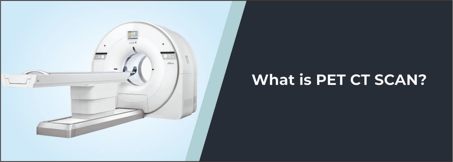
68Ga-FAPI PET- CT involves a small amount of radioactive 68Ga-FAPI tracer. The patient is positioned on the PET-CT scanner for whole-body imaging, giving a complete image of the body. PET sees areas where there is greater uptake of FAPI, and detailed anatomical information is provided through CT. Sonograms are merged to aid doctors in assessing abnormal lesions and the possible disease spread.
Common Reasons for 68Ga - FAPI PET-CT
- Detection and evaluation of cancer
- Staging of malignant disease
- Detection of metastatic lesions
- Evaluation of suspected recurrence
- Localization of primary or suspicious lesions
- Assessment of treatment response
- Evaluation when conventional imaging is inconclusive
- Treatment planning and follow-up
Conditions Evaluated
Solid Tumors
Helps evaluate various solid malignancies by identifying areas with increased FAPI uptake.
Metastatic Disease
Can help identify possible spread of cancer to lymph nodes, bones, and other organs.
Recurrent Cancer
Supports evaluation of suspected recurrent disease following previous cancer treatment.
Fibrotic and Tumor-Associated Lesions
FAPI imaging can demonstrate areas of fibroblast activation associated with certain tumor microenvironments and fibrotic processes.
Primary Tumor Localization
May assist in identifying suspicious primary lesions and assessing their extent when conventional imaging provides limited information.
Who Should Consider This Scan?
Your doctor recommends a 68Ga-FAPI PET-CT Scan if you:
- Have suspected or confirmed cancer
- Need staging of a malignant disease
- Have suspected recurrent or metastatic disease
- Require evaluation of an unknown or difficult-to-localize primary lesion
- Have inconclusive findings on conventional imaging
- Need assessment for treatment planning or follow-up
Benefits of 68Ga - FAPI PET-CT
- Provides advanced whole-body functional imaging
- Helps identify FAP-expressing tumor-associated lesions
- Combines PET functional information with CT anatomy
- Helps assess possible disease spread
- Supports cancer staging and treatment planning
- Useful in selected cases where conventional imaging is inconclusive
Why Choose Molecular Diagnostics and Therapy?
- Experienced Nuclear Medicine Specialists
- Advanced PET-CT Imaging Technology
- NABL & NABH Accredited Diagnostic Centre
- Accurate and reliable reporting
- Appropriate radiation safety protocols
- Affordable and transparent pricing
- Comprehensive Nuclear Medicine, Radiology, and Pathology services under one roof
Book Your 68Ga-FAPI PET-CT
Book an appointment at Molecular Diagnostics and Therapy for a 68Ga-FAPI PET-CT. Our nuclear medicine physicians offer state-of-the-art functional and anatomic imaging for the assessment of several types of cancer, metastatic lesions, suspicious lesions involving the primary tumor, recurrent disease, and other conditions that demand specific PET-CT evaluation.
Test information: Fasting is not required.
Reporting: Within 24 hours.
- Fasting is generally not required for a 68Ga-FAPI PET-CT scan. Follow any specific preparation instructions provided by the Nuclear Medicine team.
- Maintain adequate hydration before the examination unless your doctor has advised fluid restriction.
- Inform the Nuclear Medicine team about all current medicines, ongoing treatments, and relevant medical conditions.
- Do not stop or change any prescribed medicine unless specifically instructed by your doctor.
- Inform the team if you are pregnant, may be pregnant, planning pregnancy, or breastfeeding.
- Bring previous PET-CT, CT, MRI, biopsy, laboratory, and other relevant medical reports, if available.
- Inform the team about previous cancer treatment, including surgery, chemotherapy, radiotherapy, immunotherapy, or targeted treatment.
- Follow all hydration, medication, and other preparation instructions provided for your specific scan protocol.
Talk to Health Expert
Health Articles