68Ga-PSMA PET-CT
₹24000
Book your 68Ga-PSMA PET-CT Scan at Molecular Diagnostics and Therapy, an NABL & NABH-accredited diagnostic centre that offers advanced Nuclear Medicine, Radiology, and Pathology services. This specialized PET-CT scan employs Gallium-68 PSMA (68Ga-PSMA) to detect lesions that express Prostate-Specific Membrane Antigen (PSMA) and is special because it is used in cancer diagnosis, especially when looking at prostate cancer. PET shows how they are working. Our specialized nuclear medicine physicians employ cutting-edge PET-CT technology under proper radiation safety protocols for accurate scanning.
About 68Ga-PSMA PET-CT
The 68Ga-PSMA PET-CT Scan is a specialized nuclear medicine examination mainly used for the evaluation of prostate cancer. The 68Ga–PSMA tracer binds to cell types that express PSMA and is detected on the PET scan. It offers information on both function and anatomy, helping physicians to assess disease extent.
What is a 68Ga-PSMA PET-CT?
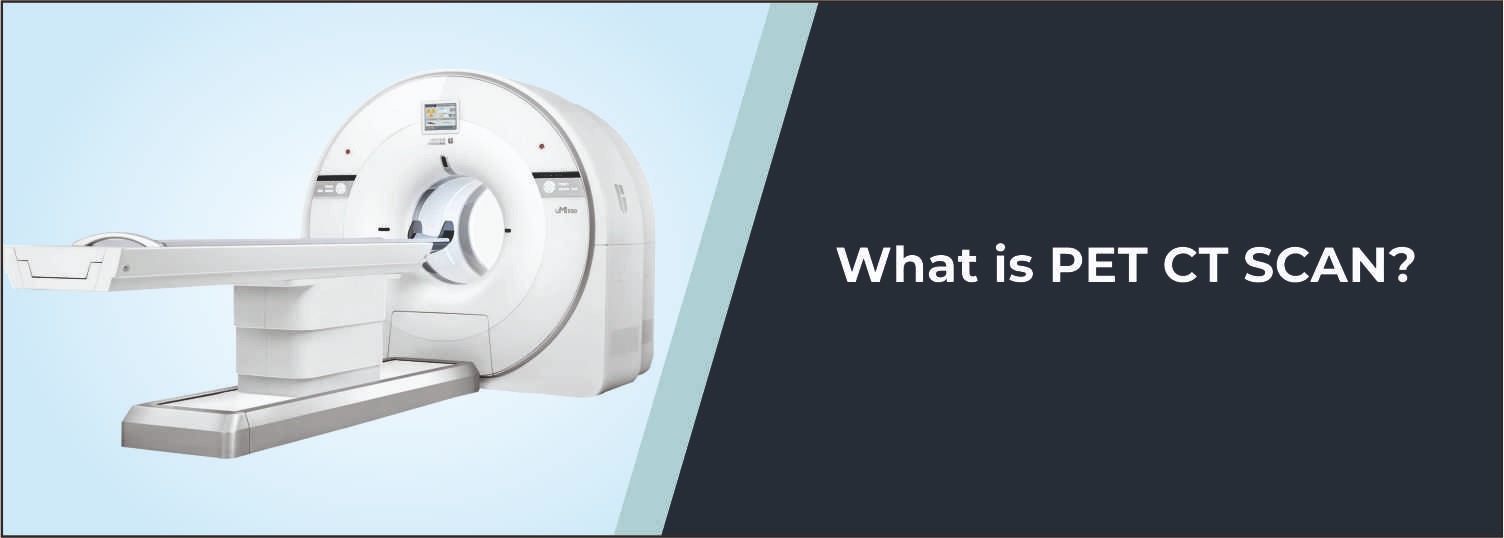
During a 68Ga-PSMA PET-CT scan, a small dose of the radioactive tracer, 68Ga-PSMA, is injected. The patient lies in the PET-CT machine for the full-body scan. The PET component creates a high-energy light beam onto the prostate to look for increased PSMA activity, and the CT component is used to get anatomical information. The two images, when combined, aid doctors in determining the location and extent of prostate cancer.
Common Reasons for 68Ga-PSMA PET-CT
- Detection of prostate cancer
- Staging of prostate cancer
- Evaluation of suspected recurrence
- Detection of metastatic disease
- Assessment of rising PSA levels
- Localization of PSMA-positive lesions
- Treatment planning
- Follow-up after prostate cancer treatment
Conditions Evaluated
Prostate Cancer
Helps detect and localize PSMA-expressing prostate cancer lesions.
Recurrent Prostate Cancer
Supports evaluation of suspected recurrence, particularly when PSA levels rise after treatment.
Metastatic Prostate Cancer
Helps identify the spread of prostate cancer to lymph nodes, bones, and other sites.
Lymph Node Involvement
Can help detect PSMA-positive lymph node metastases for accurate staging.
Bone Metastases
Helps identify PSMA-expressing prostate cancer deposits in the bones.
Who Should Consider This Scan?
Your doctor may recommend a 68Ga-PSMA PET-CT Scan if you:
- Have suspected or confirmed prostate cancer
- Need staging before treatment
- Have rising PSA after treatment
- Have suspected recurrent disease
- Need evaluation for metastatic prostate cancer
- Require assessment of PSMA-positive lesions
Benefits of 68Ga-PSMA PET-CT
- Provides whole-body functional imaging
- Helps detect PSMA-expressing lesions
- Combines PET functional information with CT anatomy
- Helps assess disease spread
- Supports staging and treatment planning
- Useful for evaluating suspected recurrence
Why Choose Molecular Diagnostics and Therapy?
- Experienced Nuclear Medicine Specialists
- Advanced PET-CT Imaging Technology
- NABL & NABH Accredited Diagnostic Centre
- Accurate and reliable reporting
- Appropriate radiation safety protocols
- Affordable and transparent pricing
- Comprehensive Nuclear Medicine, Radiology, and Pathology services under one roof
Book Your 68Ga-PSMA PET-CT
Book your 68Ga-PSMA PET-CT scan at Molecular Diagnostics and Therapy. The functional and anatomical imaging capabilities of our nuclear medicine team facilitate evaluation of prostate cancer, recurrent disease, lymph node involvement, bone metastases, and other PSMA-expressing lesions.
Test information: Fasting is not required.
Reporting: Within 24 hours*
- Fasting may be required depending on the scan protocol. Follow the fasting instructions provided by the Nuclear Medicine team.
- Stay adequately hydrated before and after the scan unless otherwise advised by your doctor.
- Inform the Nuclear Medicine team about your current medications, ongoing treatments, and relevant medical conditions.
- If you are pregnant, may be pregnant, or are breastfeeding, inform the Nuclear Medicine team before the scan.
- Carry previous PSA reports, PET-CT scans, CT/MRI reports, biopsy reports, treatment records, or other relevant medical documents, if available.
- Do not stop or change any prescribed medication unless specifically advised by your doctor.
- Follow all instructions regarding food, fluids, medications, and activity provided before the examination.
- Follow the radiation-safety and post-scan instructions provided by the Nuclear Medicine team.
Talk to Health Expert
Health Articles