CT Cystogram
₹2500
Book your CT Cystogram test at Molecular Diagnostics and Therapy – a diagnostic centre that offers highly advanced services in the field of Radiology, Nuclear Medicine, and Pathology, certified by NABL and NABH. A CT Cystogram is a special type of imaging test performed to assess the urinary bladder and other tissues to see if there is damage, leakage, or perforation of the bladder. It uses a computed tomography (CT) scan with a dye placed in the bladder to create detailed images of the bladder wall and the other structures in the pelvis. We are equipping our radiology staff with cutting-edge CT technology and proper imaging procedures to aid in the safe and accurate evaluation.
About CT Cystogram
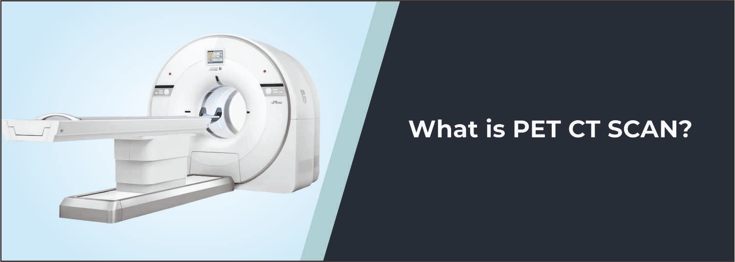
CT Cystogram is a special type of CT scan to evaluate the urinary bladder. It is especially helpful in situations where there is a medical suspicion of the bladder being damaged, leaking, or disrupted. As part of Molecular Diagnostics and Therapy, a detailed CT cross-sectional view is done to assess the bladder and other pelvic structures. Results are compared to other relevant clinical data, previous investigations, and symptoms of the patient.
What is a CT Cystogram?
In a CT cystography, a suitable contrast is inserted into the urinary bladder through a urinary catheter. After the bladder is sufficiently filled to fulfill the examination protocol, computed tomography (CT) images are taken. The contrast exposes the interior of the bladder, enabling an evaluation of the bladder wall and any leakage of the contrast into the surrounding tissues. Further CT scans are acquired before, during, or after filling the bladder based on clinical indication and examination procedure.
Common Reasons for CT Cystogram
- Evaluation of suspected bladder injury
- Detection of bladder perforation or rupture
- Assessment of suspected urinary leakage
- Evaluation following pelvic or abdominal trauma
- Assessment after certain surgical procedures
- Evaluation of postoperative bladder complications
- Investigation of abnormalities involving the urinary bladder
- Further assessment when other imaging findings require clarification
Conditions Evaluated
Bladder Injury
CT Cystogram can help identify injuries to the bladder, particularly following trauma or surgical procedures.
Bladder Perforation
The examination can help detect a breach in the bladder wall by demonstrating leakage of contrast outside the bladder.
Urinary Leakage
Contrast-enhanced bladder imaging may help identify the location and extent of suspected urine leakage.
Traumatic Bladder Injury
CT Cystogram can be useful in evaluating suspected bladder damage associated with pelvic or abdominal trauma.
Postoperative Complications
The examination may be performed when there is concern about bladder healing or urinary leakage following surgery.
Who Should Consider This Scan?
Your doctor may recommend a CT Cystogram if you:
- Have suspected bladder injury
- Have experienced pelvic or abdominal trauma
- Have symptoms or findings suggesting urinary leakage
- Require evaluation after bladder or pelvic surgery
- Have an abnormal finding requiring detailed bladder assessment
- Need further evaluation of a suspected bladder perforation
Benefits of CT Cystogram
- Provides detailed cross-sectional images of the urinary bladder
- Helps detect bladder wall injuries and contrast leakage
- Provides information about surrounding pelvic structures
- Useful in evaluating traumatic and postoperative bladder complications
- Can help identify the location of suspected urinary leakage
- Combines bladder contrast assessment with detailed CT imaging
Why Choose Molecular Diagnostics and Therapy?
- Experienced Radiology Professionals
- Advanced CT Imaging Technology
- NABL & NABH Accredited Diagnostic Centre
- Appropriate contrast administration and imaging protocols
- Accurate and reliable reporting
- Patient-focused diagnostic care
- Comprehensive Nuclear Medicine, Radiology, and Pathology services under one roof
Book Your CT Cystogram
Looking for a CT Cystogram, CT Cystogram Cost, or a reliable diagnostic centre for detailed urinary bladder imaging? Book your test at Molecular Diagnostics and Therapy. Our radiology team provides CT-based evaluation of the urinary bladder to support the diagnosis of suspected bladder injury, perforation, urinary leakage, and other conditions requiring specialised bladder imaging.
Test information: Fasting is not required.
Reporting: Within 24 hours.
- Follow the preparation instructions provided by your doctor or radiology team according to the reason for the examination.
- Fasting is not always required, but follow any fasting instructions given by the diagnostic centre.
- Inform the medical team if you are pregnant, planning pregnancy, or there is a possibility of pregnancy.
- Inform the team about any known allergy or previous reaction to contrast media.
- Inform your doctor about kidney problems, urinary tract conditions, recent surgery, or other relevant medical history.
- Provide details of current medicines and ongoing treatments.
- Inform the team if you have difficulty passing urine or any other significant urinary symptoms.
- Bring previous CT scans, ultrasound reports, X-rays, surgical records, or other relevant medical documents if available.
- A urinary catheter is generally required for administration of contrast into the bladder; inform the medical team about any previous difficulty or discomfort associated with catheterisation.
- Follow all instructions provided by the healthcare team during bladder filling and imaging.
Talk to Health Expert
Health Articles