CT Gastrography
₹6000
Book your CT Gastrography at Molecular Diagnostics and Therapy, an NABL- and NABH-accredited diagnostic centre that offers advanced diagnostic services. The specialised CT scan used to examine the stomach and the surrounding gastrointestinal tissues is called CT gastrography. This scan gives information on the stomach wall, the inside of the stomach, and surrounding organs as well as the surrounding tissues. Our experienced radiology professionals follow suitable imaging protocols to obtain detailed anatomical information for diagnosis and further clinical management.
About CT Gastrography
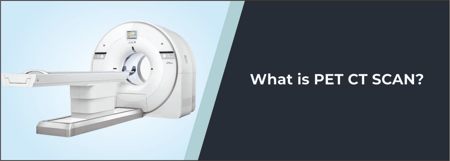
Computed tomography (CT) Gastrography is a specialized computed tomography examination that takes a cross-sectional image of the stomach and upper GI region. It is useful in the assessment of abnormalities related to the stomach wall, adjacent abdominal structures, and surrounding tissues. Molecular Diagnostics and Therapy uses advanced CT technology to get multiple detailed images from various parts of the abdomen. These projections are reformatted in a variety of planes and assist the radiologist in assessing the surrounding structures and stomach.
What is a CT Gastrography?
A CT gastrography is a CT scan of the stomach that takes place after thorough preparation and inflation of the stomach. Depending on the exam and the need, the patient has to consume the contrast orally or another contrast before the scan. The resultant images aid the radiologist in assessing the stomach wall, lumen, surrounding tissues, and other abdominal structures.
Common Reasons for CT Gastrography
- Evaluation of suspected stomach abnormalities
- Assessment of stomach wall thickening
- Evaluation of suspected gastric masses or lesions
- Assessment of gastrointestinal symptoms when detailed imaging is required
- Evaluation of suspected inflammation or other structural abnormalities
- Assessment of surrounding abdominal structures
- Further evaluation of findings seen on other investigations
- Pre-treatment or treatment planning when clinically indicated
- Follow-up evaluation of known gastric abnormalities
Conditions Evaluated
Gastric Masses and Lesions
CT Gastrography can provide detailed anatomical information about suspected masses or lesions involving the stomach.
Stomach Wall Abnormalities
The examination can help assess changes in the thickness, contour, and appearance of the stomach wall.
Gastric Tumours
CT imaging may assist in evaluating suspected gastric tumours and assessing their extent and relationship with surrounding structures.
Inflammatory Changes
The examination may help identify structural changes associated with certain inflammatory conditions affecting the stomach.
Surrounding Abdominal Structures
CT images can also provide information about nearby organs, tissues, and lymph nodes when included in the examination field.
Who Should Consider This Scan?
Your doctor may recommend a CT Gastrography if you:
- Have persistent gastrointestinal symptoms requiring detailed imaging
- Have a suspected abnormality involving the stomach
- Have an abnormal finding on endoscopy or another imaging examination
- Require evaluation of a suspected gastric mass or lesion
- Need assessment of stomach wall abnormalities
- Require further evaluation or follow-up of a known gastric condition
Benefits of CT Gastrography
- Provides detailed cross-sectional images of the stomach
- Helps assess the stomach wall and lumen
- Provides information about surrounding abdominal structures
- Supports evaluation of suspected gastric lesions and masses
- Allows multiplanar image reconstruction for detailed assessment
- Can assist with diagnosis, treatment planning, and follow-up
Why Choose Molecular Diagnostics and Therapy?
- Experienced Radiology Professionals
- Advanced CT Imaging Technology
- NABL & NABH Accredited Diagnostic Centre
- Appropriate contrast and imaging protocols
- Detailed anatomical evaluation
- Careful and reliable reporting
- Comprehensive Nuclear Medicine, Radiology, and Pathology services under one roof
Book Your CT Gastrography
Looking for a CT Gastrography, CT Gastrography Cost, or a reliable diagnostic centre for detailed stomach imaging? Book your examination at Molecular Diagnostics and Therapy. Our radiology team provides advanced CT-based evaluation of the stomach and surrounding abdominal structures to support the assessment of suspected gastric abnormalities, lesions, masses, and other conditions requiring detailed imaging.
Test information: 4–6 hours fasting is required before the examination.
Reporting: Within 24 hours*
- Follow the specific preparation instructions provided by your doctor or radiology team before the examination.
- Fasting may be required before CT Gastrography, depending on the examination protocol.
- You may be asked to drink an oral contrast preparation or another suitable preparation before the scan as instructed.
- Inform the medical team about any known allergies or previous reactions to contrast media.
- Inform your doctor if you have kidney problems, diabetes, gastrointestinal disease, or any other relevant medical condition.
- Inform the medical team about all current medicines and ongoing treatments.
- Inform the team if you are pregnant, planning pregnancy, or there is a possibility of pregnancy.
- Bring previous endoscopy, CT, ultrasound, X-ray, or other relevant gastrointestinal reports if available.
- Follow the instructions regarding stomach preparation and the timing and amount of any oral preparation provided by the diagnostic team.
- Wear comfortable clothing and remove metal objects or jewellery before the examination.
Talk to Health Expert
Health Articles