NCCT B/L SI JOINTS
₹3000
NCCT B/L SI Joints is a non-contrast computed tomography (CT) scan to obtain detailed images of both sacroiliac (SI) joints. The joints between the lower spine and the pelvis are an important interface for the transmission of load from the lower spine to the lower part of the body and are structurally known as the sacroiliac joints. CT offers detailed visualization of joint spaces and nonarticular bone structures. NCCT is an acronym for non-contrast computed tomography, and these SI joints are evaluated with the latest CT imaging technology. This examination is useful in determining changes in the joints, including erosions, sclerosis, narrowing, fracture, and other abnormalities affecting the sacroiliac joints.
About NCCT B/L SI Joints
Non-Contrast Computed Tomography (NCCT) examination is carried out without intravenous contrast, and with the help of X-rays and computerised image reconstruction, it offers detailed cross-sectional images. The complicated anatomy of the SI joints makes it possible to get all the information about the bone and joint surfaces using CT. It is indicated after a clinical examination or other imaging tests.
What is NCCT B/L SI Joints?
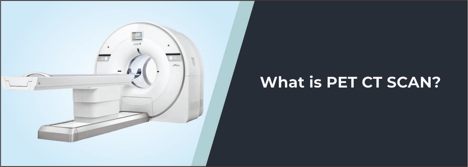
The patient is positioned properly on the CT scanning table during the examination, with scans made through the pelvis and sacroiliac joint area. The images are reconstructed into several planes for detailed images of both SI joints. This scan measures symmetry and alignment of the joints and picks up abnormalities present in them. This is a non-contrast computed tomography (CT), and routine intravenous (IV) contrast is not needed.
Common Reasons for NCCT B/L SI Joints
- Evaluation of persistent lower back or pelvic pain
- Assessment of suspected sacroiliac joint abnormalities
- Evaluation of suspected inflammatory or structural changes
- Assessment of joint-space narrowing or irregularity
- Evaluation of erosions or sclerosis
- Assessment following trauma
- Evaluation of suspected fractures involving the sacrum or pelvic region
- Follow-up of known sacroiliac joint abnormalities
- Detailed assessment when other imaging findings require clarification
Conditions Evaluated
Sacroiliitis
CT can demonstrate structural changes of the SI joints, including erosions, sclerosis, irregularity, and joint-space abnormalities that may be associated with sacroiliitis.
Ankylosing Spondylitis
The examination may help assess bony and structural changes involving the sacroiliac joints in patients being evaluated for inflammatory spinal disorders.
Degenerative Changes
CT can demonstrate bony changes and joint abnormalities associated with degenerative conditions.
Joint Structural Abnormalities
The scan can provide detailed information about joint alignment, surfaces, and surrounding bony anatomy.
Who Should Consider This Scan?
Your doctor may recommend NCCT B/L SI Joints if you:
- Have persistent lower back, buttock, or pelvic pain
- Have symptoms suggesting sacroiliac joint involvement
- Require evaluation for suspected sacroiliitis
- Have a suspected inflammatory or degenerative joint condition
- Have experienced trauma involving the pelvis or lower back
- Need detailed assessment of an abnormality seen on previous imaging
- Require follow-up of a known SI joint condition
Benefits of NCCT B/L SI Joints
- Provides detailed cross-sectional images of both SI joints
- Offers high-resolution visualization of bony structures
- Helps identify erosions, sclerosis, fractures, and structural changes
- Allows comparison of both sacroiliac joints
- Supports diagnosis and treatment planning
- Does not require intravenous contrast for routine examination
Important Preparation and Safety Instructions
- Routine NCCT generally does not require fasting unless specifically instructed.
- Wear comfortable clothing and follow positioning instructions provided by the radiology team.
- Remove metallic objects from the scanning area when requested.
- Inform the medical team about previous pelvic or spinal surgery or injuries.
- Bring previous X-rays, CT, MRI, or other relevant reports if available.
- Inform the team if you are pregnant or may be pregnant.
- Remain still during scanning to help obtain clear images.
Why Choose Molecular Diagnostics and Therapy?
- Experienced Radiology Professionals
- Advanced CT imaging technology
- Detailed musculoskeletal imaging protocols
- High-quality cross-sectional imaging
- Accurate and reliable reporting
- NABL & NABH Accredited Diagnostic Centre
- Patient-focused diagnostic services
- Comprehensive Nuclear Medicine, Radiology, and Pathology services under one roof
Book Your NCCT B/L SI Joints
If a doctor has advised detailed evaluation of both sacroiliac joints because of persistent lower back, buttock, or pelvic symptoms, NCCT B/L SI Joints provides valuable information about the joint surfaces and surrounding bones. Molecular Diagnostics and Therapy provides advanced CT imaging with appropriate professional supervision and patient-focused care.
Test information: Fasting NOT needed
Reporting: Within 24 hours*
- Routine NCCT B/L SI Joints generally does not require fasting or any special preparation unless specifically advised by the doctor or radiology team.
- Inform the radiology team about your lower back, buttock, or pelvic symptoms and any previous diagnosis involving the sacroiliac joints.
- Inform the medical team about any previous pelvic, spinal, or sacroiliac joint surgery, injury, or treatment.
- Bring previous X-rays, CT scans, MRI reports, or other relevant investigations if available.
- Wear comfortable clothing that allows proper positioning during the examination.
- Remove metallic objects from the pelvic or lower back region when requested.
- Inform the radiology team if you have any implanted medical device or metallic material in the area being examined.
- Inform the medical team if you are pregnant, planning pregnancy, or there is a possibility of pregnancy.
- Follow all positioning instructions provided by the radiology staff.
- Remain as still as possible during scanning to prevent movement-related blurring and maintain image quality.
- Inform the radiology team if pain or stiffness makes it difficult to maintain the required position.
Talk to Health Expert
Health Articles