NCCT BOTH LEGS
₹4000
Non-contrast computed tomography (CT) is an imaging technique using X-rays, with detailed cross-sectional images of both lower limbs. It offers high-resolution imaging of the bones and associated structures of both legs. It is used to diagnose fractures, traumatic injuries, bone abnormalities, and alignment issues. NCCT Both Legs is done at Molecular Diagnostics and Therapy with advanced CT imaging technology.
About NCCT Both Legs
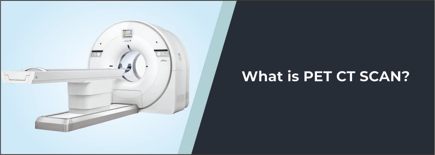
Non-Contrast CT is an X-ray study carried out without the insertion of a substance into a vein and with image reconstruction to produce detailed cross-sectional images of an X-ray. The bones of the lower limbs are very complicated, especially where joints and traumatic regions are concerned. When standard X-rays aren't enough, or alignment needs to be more precisely assessed, then CT provides more detail.
What is NCCT Both Legs?
The legs are placed in the correct position for examination at the CT, and both legs are examined. Images are obtained within the necessary regions and reassembled into detailed cross-sectional images. This scan shows dislodgement and misalignment, a fracture line, and other areas of bone damage, fragmented bones, and involvement of the joints. There is no requirement to administer intravenous contrast routinely in this examination.
Common Reasons for NCCT Both Legs
- Evaluation of suspected fractures involving both legs
- Assessment of complex or subtle bone injuries
- Evaluation following significant trauma
- Assessment of bone alignment and displacement
- Investigation of persistent or unexplained leg pain
- Evaluation of bone lesions or structural abnormalities
- Assessment of joint involvement in selected injuries
- Comparison of both legs when clinically indicated
- Preoperative planning for selected orthopaedic conditions
- Follow-up of known bone abnormalities or fractures
Conditions Evaluated
Leg Fractures
NCCT can provide detailed information about fracture location, extent, alignment, and displacement in the scanned bones.
Complex Traumatic Injuries
The examination can help evaluate complicated bony injuries following significant trauma.
Bone Lesions
Selected bone lesions and structural abnormalities can be assessed in greater anatomical detail using CT.
Joint-Related Injuries
CT can demonstrate whether a fracture or other abnormality extends into or affects an adjacent joint.
Bone Alignment Abnormalities
Detailed CT images can help evaluate the position and alignment of the bones in both lower limbs.
Who Should Consider This Scan?
Your doctor may recommend NCCT Both Legs if you:
- Have sustained injuries involving both legs
- Have persistent or unexplained pain in one or both legs
- Have suspected fractures requiring detailed assessment
- Need evaluation of complex or displaced fractures
- Require assessment of bone or joint alignment
- Have known bone abnormalities requiring follow-up
- Need comparative imaging of both lower limbs
- Require detailed imaging before selected orthopaedic procedures
Benefits of NCCT Both Legs
- Provides detailed cross-sectional images of both legs
- Offers high-resolution visualization of bony structures
- Helps identify subtle and complex fractures
- Demonstrates fracture displacement and joint involvement
- Allows comparison of corresponding structures when required
- Supports orthopaedic treatment and surgical planning
- Does not require intravenous contrast for routine examination
Important Preparation and Safety Instructions
- Routine NCCT generally does not require fasting unless specifically instructed.
- Wear comfortable clothing and follow the positioning instructions provided by the radiology team.
- Remove metallic objects from the scanning area when requested.
- Inform the radiology team about previous injuries, surgeries, or implants involving either leg.
- Bring previous X-rays, CT scans, MRI reports, or other relevant investigations.
- Inform the medical team if you are pregnant or may be pregnant.
- Remain still during scanning to help obtain clear images.
Why Choose Molecular Diagnostics and Therapy?
- Experienced Radiology Professionals
- Advanced CT imaging technology
- Detailed musculoskeletal imaging protocols
- High-quality cross-sectional imaging
- Accurate and reliable reporting
- NABL & NABH Accredited Diagnostic Centre
- Patient-focused diagnostic services
- Comprehensive Nuclear Medicine, Radiology, and Pathology services under one roof
Book Your NCCT Both Legs
If a doctor has advised detailed imaging of both legs following trauma, persistent pain, suspected fractures, or other bone abnormalities, NCCT Both Legs provides valuable information about the bones and related structures. Molecular Diagnostics and Therapy provides advanced CT imaging with appropriate professional supervision and patient-focused care.
Test information: Fasting NOT required.
Reporting: Within 24 hours*
- Routine NCCT of both legs generally does not require fasting or any special preparation unless specifically advised by the doctor or radiology team.
- Inform the radiology team about any pain, swelling, injury, fracture, or other symptoms affecting either leg.
- Inform the medical team about any previous injuries, surgeries, implants, or other treatments involving either lower limb.
- Bring previous X-rays, CT scans, MRI reports, or other relevant investigations if available.
- Wear comfortable clothing that allows both legs to be positioned easily for the examination.
- Remove metallic objects from the scanning area when requested.
- Inform the medical team if you have any implanted medical device or metallic material in either leg.
- Inform the radiology team if you are pregnant, planning pregnancy, or there is a possibility of pregnancy.
- Follow all positioning instructions provided by the radiology staff.
- Keep both legs as still as possible during image acquisition to avoid movement-related blurring.
- Inform the radiology team if pain, swelling, or an injury makes it difficult to maintain the required position.
Talk to Health Expert
Health Articles