NCCT CHEST
₹3000
Get your NCCT Chest from an NABL & NABH-accredited diagnostic centre incorporating its advanced services of Radiology and diagnostic imaging. NCCT Chest offers comprehensive, cross-sectional imaging of the lungs, airways, pleura, and other chest contents without the use of intravenous contrast material. Evaluation of lung nodules, lung infection/inflammatory lesions, interstitial lung changes, and other types of chest disease is beneficial. However, this test is sometimes recommended when a respiratory problem doesn't go away, if a chest X-ray reveals some abnormality, or to look for lung and chest disease that hasn't been diagnosed.
About NCCT Chest
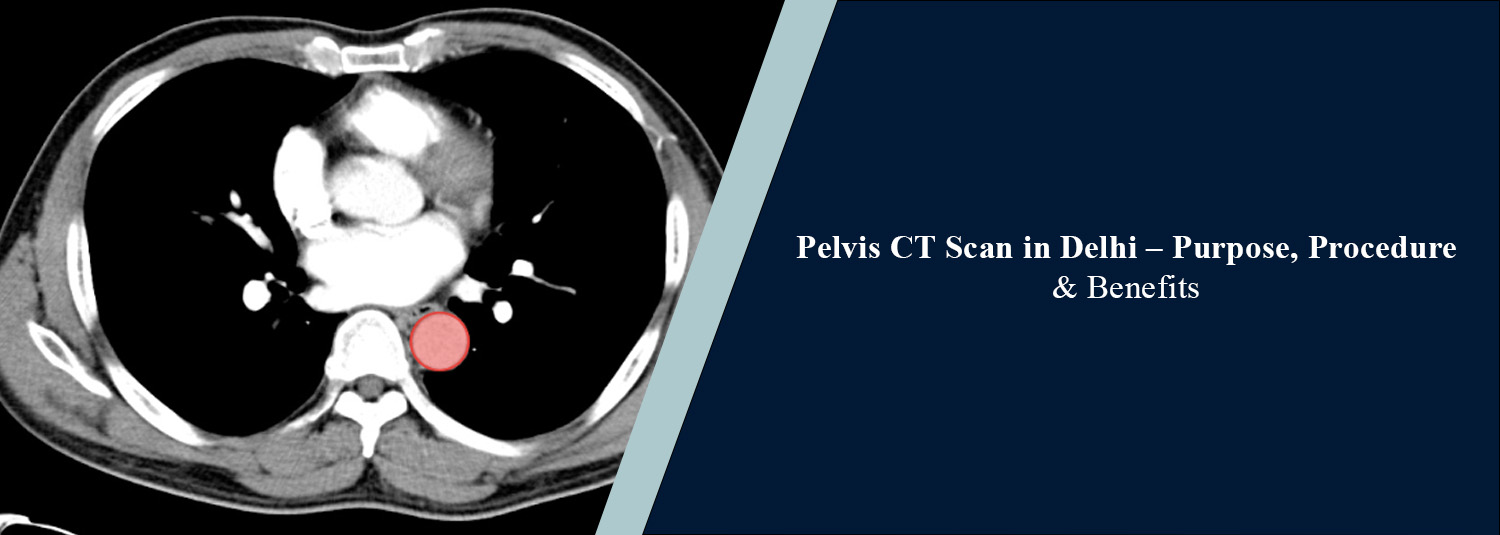
An NCCT Chest study is a special computed tomography (CT) scan that doesn't require contrast injection of a vein into your lungs or chest to get a good look at the different structures in the body. It allows detailed images to be produced that may show abnormalities that are not as clearly defined as in a conventional chest X-ray. This is an exam that can be helpful in evaluating lung tissue, airways, pleura (lining around the lung), and certain mediastinal and chest wall structures. It can be performed using a baseline chest computed tomography (CT) scan or a high-resolution technique to obtain fine detail of the lungs, with an indication for or protocol of the scan. Ongoing cough, a compromised ability to breathe, suspicion of lung infection, abnormal chest X-ray, and patients under the care of physicians for some lung conditions may undergo NCCT Chest imaging.
What is NCCT Chest?
It uses X-rays and computer processing and provides detailed cross-sectional imagery of the chest in the absence of intravenous contrast. Doctors can view the lungs and surrounding structures in various planes with the scan. It may aid in detecting pulmonary nodules, areas of inflammation and consolidation, fibrosis, emphysematous changes, abnormalities of the pleura, and other structural changes. Depending on the technique of scanning, image quality, and clinical purpose, this information may be obtained in differing ways. Further investigations may be needed to determine the cause of an abnormality.
Common Reasons for NCCT Chest
- Persistent cough: Evaluation of ongoing respiratory symptoms when further imaging is clinically indicated.
- Breathlessness: Assessment of selected lung and chest abnormalities that may contribute to breathing difficulties.
- Suspected lung infection: Investigation of pneumonia and other pulmonary changes when CT is considered appropriate.
- Abnormal chest X-ray: Further evaluation of unclear or suspicious findings on conventional radiographs.
- Lung nodules: Detection and assessment of pulmonary nodules or other focal lung abnormalities.
- Interstitial lung disease: Evaluation of suspected or previously diagnosed abnormalities affecting lung tissue.
- Tuberculosis-related changes: Assessment of selected lung abnormalities in patients being investigated for or monitored for tuberculosis.
- Emphysema: Evaluation of structural lung changes associated with chronic obstructive pulmonary disease.
- Pleural abnormalities: Assessment of pleural thickening, fluid, or other changes within the scan coverage.
- Follow-up imaging: Monitoring of selected lung nodules, infections, or other abnormalities according to the treating doctor's recommendation.
Conditions Evaluated
Lung Infections and Inflammatory Changes
NCCT chest can depict pulmonary opacities and consolidation, which are related to infection/inflammation, and ground-glass opacity. Interpretation of imaging results is performed with the symptoms, laboratory parameters, and clinical history.
Pulmonary Nodules and Masses
The scan will help to detect and identify any nodules and can help to characterise any focal abnormality in the lungs. Significance is determined by size, appearance, changes over time, and the patient's clinical risk profile.
Interstitial Lung Abnormalities
Abnormal processes affecting the lung tissue, such as specific fibrotic patterns and inflammatory interstitial lung processes, can be evaluated by CT, especially using a high-resolution protocol.
Tuberculosis-Related Lung Changes
Findings on NCCT Chest include nodules, cavities, consolidation, or scarring, which may be associated with tuberculosis. Active TB can't always be diagnosed from imaging alone, and sometimes, a series of clinical evaluation and proper laboratory testing is required to make a diagnosis.
Chronic Obstructive Pulmonary Disease and Emphysema
The exam reveals emphysematous changes and any other disorders of the lungs in structure. These can be taken into account with PFTs and clinical assessment.
Pleural Abnormalities
The involvement of pleural thickening, lesions associated with the pleura, and fluid in the pleura can be assessed with NCCT Chest. Additional testing may be advised if an apparent cause is suspected.
Airway Abnormalities
Depending on the type of scan used, the scan may be able to show selected signs and symptoms of the changes that occur in the airway walls, bronchiectasis, and mucus plugging of the bronchi and trachea.
Chest Wall and Other Thoracic Findings
On the CT scan, some abnormal areas in the chest wall, ribs, and other parts of the examination area are noted.
Why Choose Molecular Diagnostics and Therapy?
A Molecular Diagnostics and Therapy center with advanced diagnostic modules of Radiology, Nuclear Medicine, and Pathology under one roof, NABL & NABH Certified. We have the diagnostic technology to ensure that our imaging services are up to date, imaging protocols are structured, our imaging care providers are skilled, and we take a patient-first approach to imaging. We help patients through the process of the examination, proceed in an appropriate manner to ensure radiation safety, and provide quality imaging and diagnostic evaluation.
Book Your NCCT Chest
If your doctor recommended that you have an NCCT Chest, you might be able to schedule your appointment with Molecular Diagnostics and Therapy. If you consulted a doctor, bring a prescription, if available, CT scan reports if you had one, and any other medical records that are important. Let the radiology staff know that you may be pregnant before undergoing the exam and provide a history of medical reports. If you have any concerns about your pregnancy, let them know.
Test information: Usually, no fasting is required.
Reporting: Within 24 hours*
- Usually, no fasting is required before the scan.
- Inform the radiology team if you are pregnant or may be pregnant.
- Bring your doctor’s prescription, previous chest X-rays, CT scan reports, and relevant medical records, if available.
- Inform the staff about any previous chest surgery, significant medical conditions, or difficulty lying flat.
- Remove jewellery or other metallic objects from the chest area as instructed.
- Follow the radiology team’s instructions and remain still during image acquisition.
- Inform the staff if you experience discomfort or have difficulty holding your position.
- Take your regular medicines as directed by your doctor unless you have been given different instructions.
Talk to Health Expert
Health Articles