
CT Angiography Cerebral – Purpose and Risks
Talk to Health Expert

Dr. Nikunj Jain
Co-Founder and HOD - Nuclear Medicine ,MBBS, DRM, DNB, FEBNM, FANMB, Dip. CBNC
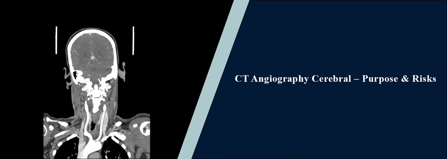
CT Angiography Cerebral, also referred
to as either cerebral CT angiography (cCTA) or brain CTA (brain CT
angiography), is an imaging method to examine blood vessels in the brain. It
allows doctors to look at the blood flow in these vessels and decide if there
is any narrowing of the vessels, blockages, aneurysms, or abnormalities. A
computed tomography (CT) scan with a dye to make detailed images of blood
vessels.
When recommended by a doctor, CT
angiography can be used at Molecular Diagnostics and Therapy to help provide
important information about the blood vessels in the brain. Patients can gain
good insight on what the test is for, how it works, and the potential risks
into it enhances their feeling to have surely measured up for it.
What Is Cerebral CT
Angiography?
Cerebral CT angiography is a special
scan of the brain that uses a high-precision CT Scan of the arteries that feed
the brain. The contrast dye is given during the test via a vein, typically in
the arm. This dye enhances the visibility of the blood vessels in the scan. A
brain CTA scan is similar to a traditional CT scan of the brain, but it
provides pictures of the blood vessels instead of the structures. It assists
physicians determine their form, size and health and alert them to problems
they might cause blood to blood flow. The test is usually fast and doesn't
involve making a surgical cut into the brain's arteries or a catheter being put
in directly.
Why Is Cerebral CT
Angiography Performed?
When doctors want to take a closer look
at the blood vessels in the brain, cerebral CT angiography may be recommended.
The test can contribute to the investigation of the following conditions:
1. Detecting Brain
Aneurysms
A brain aneurysm is a weaker spot in
the wall of a blood vessel which swells outwards. Sometimes an aneurysm may not
be felt, and sometimes it may burst and cause bleeding. Aneurysms can sometimes
be detected with a CT angiography scan, and the size and location of the
aneurysm can be determined.
2. Evaluating Stroke
It can happen when someone's blood
vessels carrying blood to the brain become blocked or if their blood vessels
start to bleed. A CT scan can also be used to find a constricted or clogged
artery and can also help the doctor determine how to treat it. It may be
combined with other imaging procedures, depending upon the symptoms and
condition of the patient.
3. Identifying
Narrowed Blood Vessels
Narrowing of the arteries can reduce
the amount of blood reaching the brain. A cerebral computerised tomography
(CTA) scan will help doctors determine the site and degree of this narrowing.
4. Detecting Blood
Vessel Abnormalities
The scan can be used to detect some
abnormalities, such as how your arteries and veins are connected in the brain
(arteriovenous malformations or AVMs). These abnormalities can impact blood
flow and sometimes even have a bleeding risk.
5. Investigating
Unexplained Symptoms
Symptoms or early test results may
suggest that the blood vessels of the brain may be an issue, and a doctor might
recommend having an imaging procedure called CT angiography. The scan may be
used as part of the evaluation when there are sudden changes in neurologic
function (neurologic status), when it is thought that the blood vessels may
have been lost, or if the headache is otherwise very serious. Based on
patients' symptoms, past medical history, and a thorough evaluation by the
doctor, the test is selected.
How Is the Cerebral
Angiography Procedure Performed?
The cerebral angiography is performed
in a diagnostic imaging centre using CT, normally. Depending on the patient's
condition and the protocol adopted by the centre, the procedure might differ.
Step 1: Preparation
Before the Scan
The staff in the radiology department
will ask you about your medical history, any medicines you are taking, your
allergies, and any reaction you've had to contrast. Let the team know whether
you suffer from kidney troubles or diabetes, or if you might be pregnant.
Removing some of the metal objects (e.g., jewelry) that may interfere with
imaging might be necessary. Don't eat, drink, or take medicines before the
scan, as recommended by the diagnostic centre.
Step 2: Positioning
on the CT Table
You will be lying on your back on the
table of your CT scan machine. The doctor will guide you into a comfortable
position with the technician, and may request that you do not move your head
during the scan.
Step 3: Injection of
Contrast Dye
An intravenous line (IV) is inserted
typically into a vein in the arm, with a contrast dye made up of iodine. The
dye circulates in the blood and enhances the visibility of the blood vessels on
the CT scans. A few people may have a small warm feeling, a metallic taste, or
they may have to pass water following the injection. Most often, these
experiences are only fleeting.
Step 4: Image Capture
A series of pictures is obtained during
the CT scan as the contrast dye passes through the blood vessels of the brain.
If directed, you will either have to hold back breathing for a few seconds or
to stay still. The imaging is a relatively rapid process, usually on the order
of several minutes, although there may be some preparation and/or other checks.
Step 5: Image Review
and Reporting
The radiographers then present a
qualified radiologist with the images, which are subsequently interpreted. The
report provides a description of how the blood vessels look and if they are
something that should be followed up with more investigation. In the event of
any concerns, your doctor will explain the results and discuss what measures to
take in the future.
What Are the Risks of
Cerebral CT Angiography?
CT angiography is commonly performed and is generally considered safe
when medically appropriate. However, like other medical imaging procedures, it
has some risks.
1. Exposure to
Radiation
The scan is performed using X-rays to
create pictures. The amount of radiation exposure is minimized in order to get
the useful information required for diagnosis. Your doctor will think about the
pros and cons before deciding to offer the scan to you.
2. Reaction to
Contrast Dye
A few patients may get nausea, itching,
a rash, or other reactions to the dye. Rare, but it can cause a severe
allergic-like reaction. Notify the medical team right away if you have any
previous reactions to contrast dye.
3. Kidney-Related
Concerns
For certain patients, particularly
those who have greatly compromised kidney function or some other risk factor,
extra caution may be needed when using iodinated contrast. Before the
procedure, you may have a check to see how well your kidneys are working.
4. Risks Related to
Pregnancy
The radiation exposure associated with
the scan requires the medical team to be notified should you be pregnant or a
woman who could be pregnant. They can determine if the exam is needed and the
options for other imaging tests.
5. Discomfort at the
Injection Site
There may be some mild discomfort,
bruising, or swelling at the intravenous insertion site. If contrast dye leaks
into the surrounding tissue, it may cause swelling or discomfort. Let the staff
know if you feel pain and/or swelling during the injection. The imaging team
takes measures to minimise the risks to you and will advise you of any special
monitoring that is required.
What Happens After
the Scan?
For the majority of patients, it's
possible to resume normal routines shortly following the treatment, unless
advised otherwise by your doctor. If indicated for your medical condition, you
will be encouraged to increase your fluid intake. Obey any guidelines the
diagnostic team suggests. Seek medical care immediately if you notice any
unusual swelling, increased pain at the injection site, difficulty breathing,
or any other red flag symptom following the scan. Talk to the doctor who is
following you for your symptoms, examination, and any other test results, and
discuss their scan report.
What Is the Cost of
Cerebral CT Angiography?
The price of a CT angiography scan of
the brain at a diagnostic center and the imaging protocol and services
performed can influence the cost of this imaging procedure. The total amount of
the test might change if other tests are ordered or medical evaluations are
ordered. Please call Molecular Diagnostics and Therapy directly before your
visit for appointment availability, preparation guidelines, and to check on the
current price.
Conclusion
CT Angiography Cerebral is a valuable imaging procedure that will enable the doctor to examine the blood vessels that supply blood to the brain. It may be useful in the diagnosis of suspected abnormal blood vessels, cerebral aneurysms, artery constriction or blockage, and stroke. The process involves radiation and an iodine-containing contrast dye and does not require surgery but is very fast and quick. If it's a medical history, advise it to the team responsible for the treatment and adhere to their guidance for a smoother and safer treatment. If the brain CTA scan has been ordered, Molecular Diagnostics and Therapy can help you to understand what it is, how you are to be prepared, and when it will be.
Frequently Asked Questions
In News
Get Our Mobile App
for Easy Access
Book tests, view reports, and manage your health records on the go. Experience convenient healthcare with Molecular Diagnostics and Therapy.
- ✔ Book tests & home collection
- ✔ View reports instantly
- ✔ Track health history
- ✔ Get notifications & reminders
- ✔ Easy appointment management
