
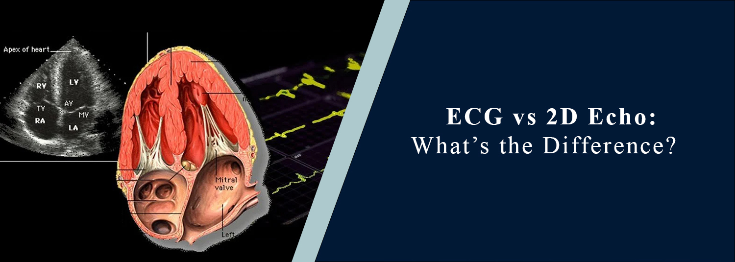
ECG vs 2D Echo: What’s the Difference?
Talk to Health Expert

Dr. Nikunj Jain
Co-Founder and HOD - Nuclear Medicine ,MBBS, DRM, DNB, FEBNM, FANMB, Dip. CBNC
Timely and correct diagnosis is often essential to avoid complications due to heart problems. Doctors usually
recommend an ECG and 2D Echo when patients experience symptoms such as chest pain, shortness of breath, palpitations, or dizziness. Both are used to assess the health of the heart, but have different functions and give different kinds of information.
Knowing the differences between these tests will help you feel a lot more confident and educated in cases when your doctor recommends them.
What Is an ECG?
An ECG or Electrocardiogram is a procedure that captures the electrical impulses of the heart. The heart obeys electrical impulses, and the test records them to confirm that the heart is working properly.
Small electrodes are put on the legs, arms, and chest during the procedure. These electrodes record the electrical responses and chart them as wave patterns. It is fast, easy, and normally done in a few minutes.
One primary use of ECG is to diagnose irregular heart rhythms, diagnose a heart attack, and track heart rate. It is a frequently recommended test in situations of sudden symptoms, as this test yields instant revelations.
What Is a 2D Echo?
A 2D Echo or 2D Echocardiography is an imaging test that involves the use of sound waves to form real-time images of the heart. A 2D Echo provides a view of the true structure and movement of the heart, as opposed to an ECG, which records electrical signals.
In this process, a gadget known as a transducer is placed on the chest. It transmits sound waves in the body, which reflect to form images on a screen. Such images assist physicians in seeing the functioning of the heart chambers, valves, and muscles.
A 2D Echo is particularly viable to find structural issues, determine the functioning of the heart valve, and determine the efficiency of heart pumping.
Key Differences Between ECG and 2D Echo
This comparison shows that the two tests offer various yet complementary information on the health of the heart.
When Is an ECG Recommended?
The initial procedure in assessing the conditions of the heart is usually by ECG. It is popular both as an emergency and routine application because it is fast and simple.
The doctors can prescribe an ECG in case of:
Some of the possible causes of sudden chest pain or discomfort include:
Poor heartbeat rhythm or rapid heartbeat.
Fainting or lightheadedness
Suspected heart attack
Regular examinations of persons at risk or factors such as high blood pressure or diabetes.
Due to its fast outcomes, ECG is a very vital tool in the detection of cardiac problems at an early stage.
When Is a 2D Echo Recommended?
A 2D Echo is recommended in case a more detailed inspection of the heart is required. It is commonly done as a follow-up to an ECG in case of abnormalities revealed or persistent symptoms.
Should some suspicions arise on unexplained breathlessness, the possibility of valve disease, or evidence of heart failure, you might be requested to have a 2D Echo. It is also applicable in assessing heart murmurs and to follow up on heart conditions already existing.
This test presents a better description of the structural functioning of the heart, and it aids in proper diagnosis and treatment planning.
Do You Need Both Tests?
Both ECG and 2D Echo are often used simultaneously in order to have a comprehensive picture of heart conditions. ECG is concerned with the heart's electrical activity, whereas a 2D Echo enhances visual information regarding the heart structure and its performance.
To take an example, an ECG may display an abnormal rhythm, but a 2D Echo has the ability to figure out whether the problem affects valves or weakened cardiac muscles. Such a combined treatment will lead to a more accurate diagnosis.
Which Test Is Better?
No test is more useful than another, as each has a specific purpose to fulfill. ECG should be used when we want to find electrical abnormalities, but 2D Echo is more appropriate when we want to diagnose the physical state of the heart.
It all depends on the symptoms and medical history of the patient. In most cases, physicians will use a combination of both tests to come up with an accurate diagnosis.
Are These Tests Safe?
ECG and 2D Echo are both non-invasive and totally safe. They are radiation inactive and do not have adverse side effects. The testing can be performed immediately, after which the patient can continue with their normal activities.
This distinguishes them as repeatable in occurrence in the event that tracking is needed across a time frame.
Conclusion
ECG and 2D Echo are vital in heart management in modern heart care, as each provides meaningful information in different ways. Although ECG assists in the identification of the problems associated with the heart rhythm and electrical activity, 2D Echo offers a clear picture of the heart structure and functioning.
The difference between these tests is known to prevent confusion and anxiety in case they are ordered. In case you have symptoms such as chest pain, shortness of breath, or irregular heartbeat, prompt testing will result in early diagnosis and improve the outcomes of the treatment process.
The combination of ECG and 2D Echo is crucial to keeping the heart healthy and avoiding severe complications, which means that there will be a more informed and proactive approach to heart care.
Frequently Asked Questions
In News
Get Our Mobile App
for Easy Access
Book tests, view reports, and manage your health records on the go. Experience convenient healthcare with Molecular Diagnostics and Therapy.
- ✔ Book tests & home collection
- ✔ View reports instantly
- ✔ Track health history
- ✔ Get notifications & reminders
- ✔ Easy appointment management
