
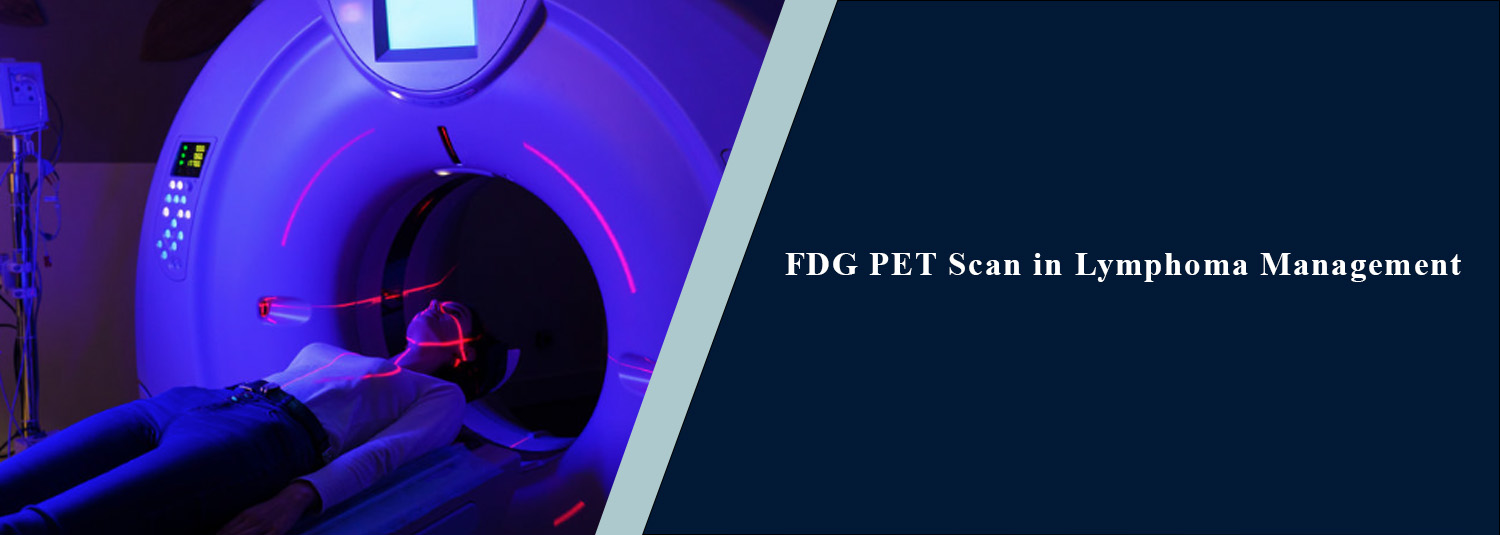
FDG PET Scan in Lymphoma Management
Talk to Health Expert

Dr. Nikunj Jain
Co-Founder and HOD - Nuclear Medicine ,MBBS, DRM, DNB, FEBNM, FANMB, Dip. CBNC
Lymphoma is a type of cancer that
develops in the lymphatic system, which plays a crucial part in the body's
immune system. It also involves organs (spleen, bone marrow, liver, or other
tissue) as well as lymph nodes. Lymphoma develops in various locations and acts
differently in each type; therefore, it is vital that it is accurately assessed
so that treatment and its response are planned for.
An FDG PET CT scan is one of the more
sophisticated types of imaging exams that uses a combination of PET and CT
scans. It gives information about the structure of tissues and about the
metabolic activity of tissues. FDG PET CT has proven to be useful in the
management of staging and response assessment in many FDG-avid lymphomas and is
now an integral part of the recommended evaluation of the FDG lymphoma patient.
What Is an FDG PET CT
Scan?
Fluorodeoxyglucose (FDG) PET CT is a
low-dose scan that involves administering a radioactive dye that mimics
glucose. The increased level of glucose consumption by many cancer cells helps
to identify areas of elevated metabolism on a PET scan. The PET component shows
increased metabolic activity, and the CT component shows the detail of anatomy.
Putting these images together helps doctors determine where the lymphoma is, as
well as how active the lymphoma is metabolically.
Why Is FDG PET CT
Used in Lymphoma?
PET CT is not just used to detect
enlarged lymph nodes in lymphoma. It gives information on how the distribution
and metabolic activity of disease occurs inside the body. In FDG-avid
lymphomas, PET CT is helpful in the initial staging process to help determine
the extent of disease. It is also used as a measure of the effectiveness of
treatment, especially following treatment. For FDG-avid lymphomas, the Lugano
classification officially includes FDG PET CT as part of routine staging and response
evaluation.
FDG PET CT for
Lymphoma Staging
Staging is important to help the doctor
understand the extent of the spread of lymphoma and plays a key role in
treatment planning. An oncology PET CT scan is used to detect metabolically
active lymphoma in various areas of the body, such as lymph nodes as well as in
certain extranodal locations. It also gives information about involvement of
other organs, such as the spleen or bone marrow, if applicable, based on the
type and clinical condition of the lymphoma. PET CT is very useful for some
non-Hodgkin lymphomas, especially for those that are aggressive or are
FDG-avid, and in Hodgkin lymphoma. Not all lymphomas, however, behave in the
identical manner on FDG PET; thus, the type of imaging depends on the type of
lymphoma and clinical circumstances.
Role of FDG PET CT
During Treatment
PET CT is also used to determine the
response of the lymphoma to therapy. An interim PET CT scan is conducted during
treatment in certain cases of lymphoma and treatment for the purposes of
diagnosis. The results offer data on metabolic response and/or are part of the
basis for treatment decisions in specific scenarios. Interim PET, however,
shouldn't be routinely ordered or evaluated for each type of lymphoma. The time
and usefulness vary with the type of disease and treatment protocol.
FDG PET CT After
Treatment
An important use of an FDG PET CT scan
is to assess response/activity at the end of treatment in many lymphomas that
respond to immunotherapies such as chemotherapy and radiation therapy
effectively. CT shows a swollen lymph node or mass that has been left over in
the area from treatment that is not actually active and represents fibrosis or
other changes. PET also offers other metabolic information which is useful in differentiating
metastases from residual non-metabolic abnormalities. In lymphomas with
FDG-avid tumours, the treatment response is routinely assessed by the Lugano
classification and the Deauville 5-point scale.
What Is the Deauville
Score?
To simplify the interpretation of FDG
PET results in lymphoma, a score on a 5-point scale has been developed: the
Deauville score. This compares the FDG uptake in regions of possible disease to
an FDG uptake activity standard from normal tissue (such as blood pool and liver).
This standardization is used to characterize the extent of response, residual
activity, or persistent/progressive disease seen on the scan. When interpreting
the score, it is essential to take the specific type of the patient's lymphoma,
treatment history, symptoms, lab results, and previous imaging into account.
How Is an FDG PET CT
Scan Performed?
The patient is given certain routine
instructions beforehand, before the scan, such as fasting. Glucose levels are
sometimes measured before administration of FDG due to the impact that the
glucose level has on the distribution of the FDG. FDG is given for a short
period, typically via an injection into the bloodstream. The patient is then
placed in the rest position for a certain amount of time, permitting the tracer
to passively diffuse throughout the body. The patient is then placed on a PET
CT scanner for imaging and then removed. The acquisition of the CT usually
takes place in the course of the same examination and is often followed or
carried out together with the acquisition of the PET image. The technique used
changes depending on patient condition and examination objectives.
What Does an FDG PET
CT Scan Show in Lymphoma?
An FDG PET CT scan can provide information about the location and
metabolic activity of lymphoma. Depending on the disease, it may help identify
involved lymph nodes, extranodal disease, and areas that require further
clinical evaluation. The scan can also be compared with previous PET CT
examinations to assess changes in metabolic activity over time. This comparison
can be particularly useful when evaluating treatment response. However, FDG
uptake is not specific to cancer. Inflammation and infection can also show
increased FDG uptake. Therefore, PET CT findings should be interpreted by an
experienced specialist along with clinical and laboratory information.
FDG PET CT Scan Test
Details
The exact FDG PET CT scan test details can vary depending on the
patient's lymphoma subtype, treatment stage, medical history, and the purpose
of the examination. Patients may be advised to avoid strenuous physical
activity before the scan and follow fasting instructions provided by the
imaging centre. Diabetic patients may require special preparation because blood
glucose levels can influence FDG uptake. Patients should inform the medical
team about diabetes, pregnancy or possible pregnancy, medications, previous
reactions to contrast material, and other relevant medical conditions before
the examination.
Is FDG PET CT Safe?
FDG PET CT involves exposure to radiation from both the radiotracer and
the CT component. The amount is carefully considered against the clinical
benefit of obtaining the necessary diagnostic information. The FDG used for PET
imaging has a relatively short physical half-life and is gradually eliminated
from the body. Patients are given specific instructions when necessary,
particularly regarding hydration and urination after the examination. As with
any medical imaging procedure, the decision to perform PET CT should be based
on the patient's clinical requirements and the recommendation of the treating
doctor.
FDG PET CT Scan
Charges
The FDG PET CT scan charges can vary between diagnostic centres
depending on the type of scan, imaging protocol, radiotracer used, location,
and additional services included in the examination. Patients should confirm
the current charges and preparation requirements directly with the diagnostic
centre before scheduling the scan. It is also important to understand what is
included in the quoted charges, particularly when additional CT contrast or
specific imaging protocols are required.
Why Choose Molecular
Diagnostics and Therapy for FDG PET CT?
Molecular Diagnostics and Therapy provides advanced nuclear medicine and
diagnostic imaging services to support cancer evaluation and treatment
monitoring. For patients undergoing lymphoma assessment, an FDG PET CT scan can
provide valuable metabolic and anatomical information when clinically
indicated. The scan should be performed according to the referring doctor's
requirements, with interpretation by qualified specialists. Using advanced PET
CT technology and standardized approaches to lymphoma response assessment can
help provide clinically useful information for oncologists and other treating
specialists.
Conclusion
In the treatment of various FDG-avid
lymphomas, FDG PET CT is emerging as a very valuable imaging modality. It is
useful for early staging, treatment response, and post-treatment evaluation.
PET images are interpreted with greater uniformity by standardized systems like
the Lugano classification or five-point Deauville scale. But PET CT is just one
component of lymphoma treatment. The results should be evaluated in conjunction
with biopsy results, clinical exam, lab work, past treatments, and other
imaging modalities.
The timing and frequency of PET CT are dependent on both the type of lymphoma and the treatment plan of each person.Molecular Diagnostics and Therapy provides state-of-the-art diagnostics and nuclear medicine services to complement a comprehensive oncology evaluation. Before scheduling your scan, discuss with the medical team the best scan protocol well suited for your lymphoma and how to prepare for the scan.
Frequently Asked Questions
In News
Get Our Mobile App
for Easy Access
Book tests, view reports, and manage your health records on the go. Experience convenient healthcare with Molecular Diagnostics and Therapy.
- ✔ Book tests & home collection
- ✔ View reports instantly
- ✔ Track health history
- ✔ Get notifications & reminders
- ✔ Easy appointment management
