
PET vs CT Scan: Differences, Uses
Talk to Health Expert

Dr. Nikunj Jain
Co-Founder and HOD - Nuclear Medicine ,MBBS, DRM, DNB, FEBNM, FANMB, Dip. CBNC
In medicine, images are valuable to
diagnose and assess the symptoms of an illness, to determine a treatment plan,
and to evaluate a patient's condition. Two commonly discussed imaging
techniques are PET and CT scans. Both give a detailed look at what's going on
inside the body but require different methods to provide different information.
A CT scan is most commonly used to
produce detailed images of the organs, bones, blood vessels, and soft tissues.
Metabolic or cellular activity is determined using a radioactive tracer in a
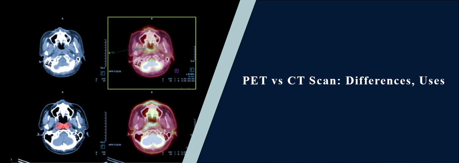
PET scan. Both types of information are obtained by a combined PET and CT scan.
It helps patients understand why a doctor might recommend one test over another
and explains the different types of scans.
What Is a CT Scan?
A computed tomography (CT) scan is an
X-ray picture of the body taken by a computer. These images are then manipulated
into various planes, different views, and three-dimensional views that enable
doctors to view internal structures up close and personal. The images are made
of various areas of the body such as the head, chest, abdomen, pelvis, spine,
bones, and blood vessels. The contrast is administered intravenously or orally,
depending on the exam, to make some tissues and blood vessels more visible
during the exam.
What Is a PET Scan?
PET refers to positron emission
tomography. Although PET has anatomical information, it is important to keep in
mind that it provides information about biological activity within tissues
rather than primarily anatomical information, as is the case for CT. PET scans
use a small amount of radioactive tracer injected into a person, typically as a
small injection. The most popular PET tracer is FDG, or a radioactive version
of glucose. On PETs, more active areas indicate high glucose uptake due to the
fact that many cancer cells have a higher glucose uptake than normal cells. PET
plays a role in various fields of medicine such as oncology, cardiology, and
neurology. It varies with the type of tracer and the question being asked in
the clinic.
PET vs CT Scan: Key
Difference
The primary difference between PET and
CT is that PET allows for measurement of information rather than CT does not.
CT primarily shows where an abnormality is and what it looks like anatomically.
PET measures the metabolic activity or metabolism of tissues. For instance, a
CT scan reveals an enlarged lymph node or a mass and gives information on the
size and location of the mass. A PET scan demonstrates the metabolic level of
that area when using a specific tracer.
PET vs CT Scan for
Cancer
The use of PET and CT is different,
with both having an important role in cancer care. CT is used to find out if
the patient has tumors, to determine the size and location of the tumors, and
to see what is nearby; it is also used to see some lymph nodes; and it is used
to plan surgery or biopsies, or other treatments. It is also employed as a
control of cancer and to search for recurrence.
PET provides information about
metabolic activity and is of value for identifying active disease in the right
forms of cancer. It is also used to find out if the disease has started anew or
if the treatment has worked. Thus, a combined PET and CT exam is a single
examination that yields anatomical and metabolic information.
What Is a PET/CT
Scan?
PET and CT allow for the imaging of the
same patient within the same session and are together known as a PET-CT scan.
The CT part gives you anatomical information; the PET part gives you functional
or metabolic information. Combining the two images can assist the doctor in
locating an area of abnormal functioning and more precisely determine where an
abnormal area is located in the body. This has the potential to provide
significant information in an oncology setting and impact diagnosis, extent of
disease, treatment planning, and/or follow-up.
CT Scan Test: When Is
It Used?
There are a lot of medical conditions
that require a CT scan. It is used to assess injuries, infections, tumors,
bloody conditions, blood vessel abnormalities, kidney stones & bladder
stones, lung disease, and various other conditions. As a tool in cancer management,
CT is used to help identify tumors and characterize them, evaluate the extent
of disease, plan procedures, plan treatment, and assess response and/or
recurrence. The type of CT that is performed varies according to the region of
the body being examined and the question being asked. Contrast is needed for
certain tests.
CT Scan Procedure:
What Happens?
A person is on a motorised table that
is moved during a CT scan procedure. The CT machine delivers several images
from a variety of angles obtained from a rotating X-ray machine. During image
acquisition, the patient is generally required not to move. Some X-rays of the
CT scans require contrast dye: it is swallowed or put in a vein. CT scans
typically take a few seconds, and the length of time varies depending on the
area and procedure being used.
How Is a PET Scan
Performed?
A PET exam involves the use of a
radiotracer that is injected into the patient and typically a period of quiet
during which the radiotracer circulates throughout the body. The discontinuance
is meant so that the tracer builds up in the tissue that is being assessed. The
patient then lies on a PET/CT bed as images are captured. The quality of images
is affected by motion; therefore, it is important to remain still while being scanned.
The preparation varies based on the tracer and kind of PET exam. Fasting and
blood glucose management could be of special significance for FDG PET, since
there is a dependence of the distribution of the tracer on blood glucose.
Which Scan Is More
Accurate: PET or CT?
There is no universal answer to which scan is more accurate because PET
and CT measure different things. CT is highly useful for detailed anatomical
assessment, while PET can provide metabolic information that may reveal active
disease not fully characterized by anatomy alone. In some clinical situations,
one test may be more appropriate than the other. For selected cancer
evaluations, PET/CT can provide complementary information from both techniques
and may offer a more comprehensive assessment than either examination alone.
Therefore, CT scan accuracy should be considered in relation to the specific
condition, body part, scan protocol, image quality, and clinical question
rather than as a single universal number.
PET vs CT Scan:
Radiation Exposure
Both PET and CT involve radiation, but the sources are different. CT
uses X-rays to create images. PET uses a radioactive tracer that emits
radiation detected by the PET scanner. A combined PET/CT therefore involves
radiation from both components. The radiation exposure is carefully considered
against the expected medical benefit of the examination. Doctors recommend
these tests when the information they provide is clinically useful.
Which Scan Should You
Get?
The choice between PET, CT, or PET/CT depends on the reason for imaging.
A doctor may recommend CT when detailed anatomical information is the primary
requirement. PET may be preferred when information about tissue metabolism or
cellular activity is important. PET/CT may be recommended when both anatomical
and functional information are needed. The choice can depend on the suspected
disease, symptoms, previous test results, treatment history, and the specific
clinical question.
PET and CT Imaging at
Molecular Diagnostics and Therapy
Molecular Diagnostics and Therapy provides advanced diagnostic imaging
and nuclear medicine services to support the evaluation and monitoring of
various medical conditions. When a PET, CT, or PET/CT examination is
recommended, the appropriate imaging protocol can be selected according to the
patient's clinical requirements. The images are interpreted by appropriately
trained specialists, and the findings are provided to the referring doctor for
further clinical decision-making.
Conclusion
Both PET and CT scans are types of
imaging, and they have their unique advantages. CT yields detail of anatomy;
PET yields detail of metabolism and cells. When functional information is
clinically needed, the two technologies are combined in a PET/CT scan and lead
to a more complete picture.
A computed tomography scan is useful for imaging tumors, injuries, organs, blood vessels, and numerous other conditions; and PET has a significant role in specific cancer, neurologic, and cardiac testing. The correct test should be decided on the basis of the symptoms, medical history, and the doctor's recommendation for the patient. Advanced imaging and nuclear medicine techniques are available at Molecular Diagnostics and Therapy to help with accurate diagnosis and evaluation of treatment in individual cases.
Frequently Asked Questions
In News
Get Our Mobile App
for Easy Access
Book tests, view reports, and manage your health records on the go. Experience convenient healthcare with Molecular Diagnostics and Therapy.
- ✔ Book tests & home collection
- ✔ View reports instantly
- ✔ Track health history
- ✔ Get notifications & reminders
- ✔ Easy appointment management
