NCCT KUB
₹3000
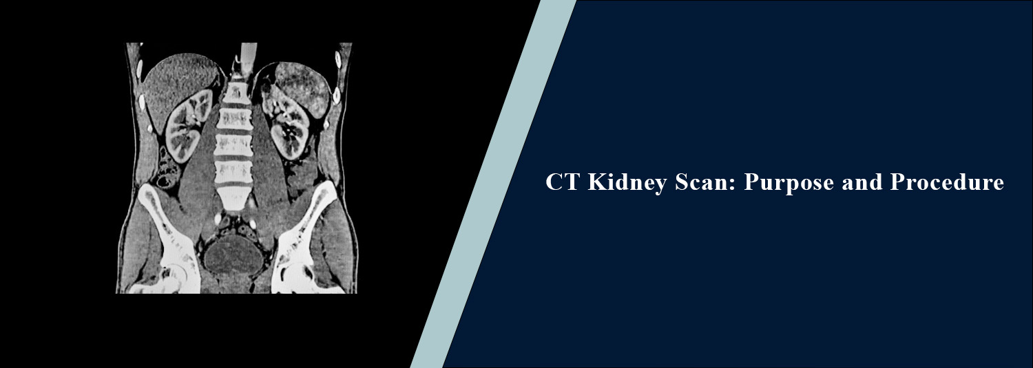
Book your NCCT KUB CT Scan at Molecular Diagnostics and Therapy, an NABL & NABH-accredited diagnostic centre with a dedicated team of diagnosticians, which provides state-of-the-art radiology and CT scans for detailed examination of the kidneys, ureters, and bladder. NCCT KUB is an abbreviation for Non-Contrast Computed Tomography of the Kidneys, Ureters, and Bladder. The NCCT KUB test is a type of CT scan that helps identify stones, blood in the urine, low back pain, and bladder issues. The NCCT KUB test can produce high-resolution images that can reveal even small stones, determine their location and size, and detect other abnormalities that can cause pain and urinary problems. From radiology specialists to technology to safe and accurate reporting.
About NCCT KUB
NCCT KUB is an imaging test that is performed for a thorough examination of the entire urinary system. These structures include the urinary bladder, ureters, and kidneys. Mostly, this test is performed when the presence of a stone in the urinary system or other diseases that may be responsible for sudden or serious urinary symptoms is suspected.
What is NCCT KUB?
Non-contrast computed tomography of the KUB, which is done without IV contrast. NCCT KUB has developed into a common technique used to diagnose stones in the urinary tract. This study can also give information about the size, number, location, and density of stones, as well as determine the presence or absence of obstruction.
Common Reasons for NCCT KUB
Flank or Side Pain
NCCT KUB may be recommended for sudden or persistent pain in the side, lower back, or abdomen when a urinary stone is suspected.
Suspected Kidney Stones
Most doctors will tell you this whenever the patient exhibits signs of having stones in the kidney or urinary tract.
Blood in Urine
The test can help rule out blood in the urine and find abnormalities in the urinary tract that could be related to the symptom.
Painful Urination
NCCT KUB may be taken into consideration when there are urinary symptoms and the condition is thought to be a stone or a blockage.
Recurrent Urinary Stones
It helps to identify stone recurrence as well as identifying the location of the stone in case it needs to be removed.
Urinary Obstruction
A stone or other structural abnormality may be blocking your flow of urine. An NCCT KUB may detect this.
Follow-Up of Known Stones
It can be performed to follow known urinary calculi and to evaluate any change in their position or size.
Conditions Evaluated
Kidney Stones
An NCCT KUB can reveal the stone's precise appearance and assist in ascertaining the size, quantity, location, and density of the stone.
Ureteric Stones
It can also detect stones in the ureters and determine the stone's location and effect on urine flow.
Urinary Bladder Stones
NCCT is capable of demonstrating calculi within the urinary bladder along with their size and location.
Hydronephrosis
The presence of a stone in the urinary tract can cause blockage and affect the kidney. This is detected on the NCCT KUB.
Ureteric Obstruction
The test is used to rule out obstruction or narrowing of the ureter and to see if it is affected by a stone.
Urinary Tract Abnormalities
There may also be structural abnormalities of the kidney, ureters, bladder and other tissues of the urinary tract.
Other Abdominal and Pelvic Findings
As the procedure encompasses the relevant abdominal and pelvic area, other abnormalities may be revealed within neighbouring structures during the investigation.
Why Choose Molecular Diagnostics and Therapy?
Molecular Diagnostics and Therapy is a NABL & NABH Accredited Diagnostic Centre with a state-of-the-art CT and Radiology Centre, a team of experienced radiology specialists, and high-end imaging technology for diagnostic excellence. Our centre adheres to the required safety norms with an emphasis on reliability and promptness of reporting. With a commitment to patient-centricity, transparency, and affordable diagnostic service, Molecular Diagnostics and Therapy helps patients and referring physicians with reliable Imaging Data.
Book Your NCCT KUB
For NCCT KUB at Molecular Diagnostics and Therapy, patients can contact the centre to confirm examination details and appointment availability. The examination is performed according to the clinical indication and symptoms being evaluated.
Test information: Usually, no fasting is required.
Reporting: Within 24 hours*
- Usually, no fasting is required before an NCCT KUB.
- Follow the centre's instructions regarding water intake before the examination. Do not intentionally overfill or empty the bladder unless instructed.
- Inform the radiology team about any previous surgery, procedure, injury, or treatment involving the kidneys, ureters, bladder, or urinary tract.
- Inform the centre about any known kidney stones, urinary obstruction, urinary tract abnormalities, or previous stone procedures.
- Tell the radiology team if you are pregnant or planning pregnancy, as CT uses ionising radiation.
- Inform the radiology team about your current medicines and relevant medical conditions.
- Bring previous CT scans, X-rays, ultrasound reports, MRI reports, or other relevant urinary and abdominal investigations if available.
- Remove jewellery, belts, or other unnecessary metallic objects before the examination.
- Wear comfortable clothing and follow the positioning instructions provided by the radiology team.
- Inform the staff if you have severe abdominal, flank, or back pain or difficulty maintaining the required position.
- Keep the body still during scanning to reduce motion-related image degradation.
- Follow any additional instructions provided by the radiology team based on your clinical condition.
Talk to Health Expert
Health Articles