
Common Conditions Diagnosed Using SPECT Imaging
Talk to Health Expert

Dr. Nikunj Jain
Co-Founder and HOD - Nuclear Medicine ,MBBS, DRM, DNB, FEBNM, FANMB, Dip. CBNC
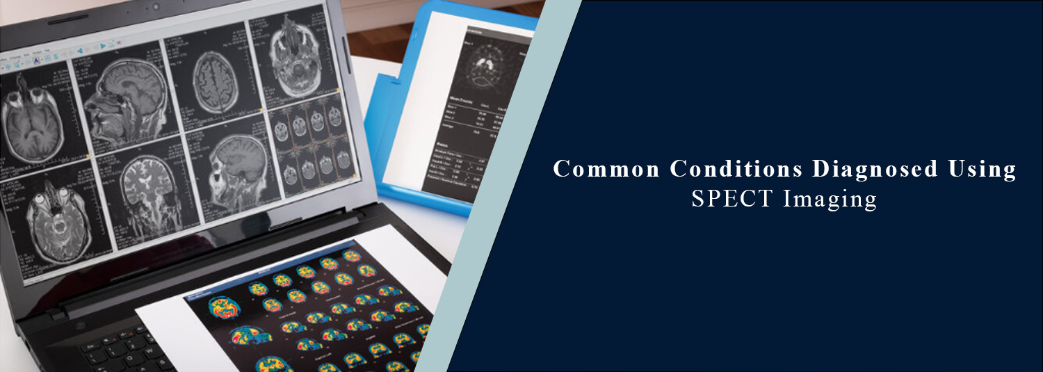
SPECT is a type of nuclear medicine imaging that is used to
determine the blood flow and function of various organs, tissues, or bones. A
SPECT scan, as opposed to certain imaging tests that mainly reveal the
structure of the body, can supply info regarding function within the body. It
is beneficial in assessing a number of heart, bone, and neurological disorders.
SPECT CT scan also uses computed tomography to provide doctors
with detailed information about the anatomy of the body as well as functional
information from a SPECT scan. Depending on the patient's symptoms and medical
history, a cardiac SPECT, a bone SPECT, or either of the other types of SPECT
examination is recommended. The high level of nuclear medicine and diagnostic
imaging services at Molecular Diagnostics and Therapy ensures accurate
diagnoses and treatment planning.
What Is SPECT Imaging?
Single Photon Emission Computed Tomography (SPECT) is a type of
PET scan. A radiological modality employing a radioactive tracer in very small
amounts in order to evaluate function within the body. The tracer is
administered to the patient and goes into the region of the body under study.
The tracer emits radiation, which is registered by a special camera, and images
of tissue or organs are formed to depict the activity of the selected tissue or
organ. SPECT can provide functional information, thus allowing identification
of abnormalities that can sometimes not be readily seen on the structural
images.
What Is a SPECT CT Scan?
A SPECT CT scan is a combination of two imaging modalities in one!
SPECT is a means of providing information on biological activity, whereas CT is
a means to provide detailed information about body structures. These images
allow the doctor to localise a lesion more precisely (and determine its
anatomical location better) when they are correlated. This is especially
helpful in studying some of the medical conditions of bones, the heart, and
many others.
Cardiac SPECT for Heart Conditions
One of the most frequent uses of SPECT is for the imaging of the
heart. The use of cardiac SPECT is primarily for assessment of blood flow to
the heart muscle. Cardiac SPECT can be used to diagnose a heart attack if a
patient has suspected CAD or symptoms like chest pain or unexplained shortness
of breath. Can assist in locating regions of the heart that are not receiving
enough blood. Cardiac SPECT is also used to determine the extent of an existing
heart disease and to find out how efficiently the heart muscle is working
during stress.
Coronary Artery Disease
CAD is the narrowing or blockage of blood vessels that carry blood
to the heart. This can help restrict blood supply to the heart muscle. When
used in conjunction with a cardiac SPECT scan, these areas can become apparent
in the presence of decreased blood supply. The results are then compared with
the patient's physical exam, symptoms, medical history, and other heart tests
for interpretation. Results can aid physicians in determining if more
investigations and/or treatment is necessary.
Bone Conditions and Bone SPECT
Bone SPECT can be used to investigate abnormal activity in bones.
It is recommended for patients who have any nerve-racking bone pain or when
another imaging test notes an area that should be investigated further. Bone
SPECT might be beneficial for evaluating some stress injuries and fractures,
infections, arthritis, and other abnormalities of bone. If bone SPECT is used
along with CT, it is often possible to see more accurately where the abnormal
area is located.
Stress Fractures and Difficult-to-Detect
Injuries
It may not be obvious which bones have a small bone injury or
stress fracture on the initial X-ray. A bone scan or bone SPECT can find an
increased uptake in the affected area. This can be useful for those who suffer
from persistent pain but may not have a definite diagnosis from their initial
imaging test. Based on what is being suspected to have happened, as well as the
individual's condition, the doctor may decide on one type of test over another:
SPECT, CT, or MRI.
Bone Infection
An infection may affect the bone and cause an abnormal movement of
the bone. A special test, SPECT or SPECT CT, is performed sometimes when
additional information is needed about an infected bone. The imaging findings
are analyzed and considered along with the symptoms, blood tests, physical
exam, and other tests that are done before a diagnosis is made.
Arthritis and Joint Problems
In certain joint and bone diseases, SPECT imaging can also be used
to assess the condition. Regions of heightened activity might be linked to
inflammation or degeneration, injury, or other abnormalities. SPECT CT is of
benefit in patients who have pain involving more than one area, where the
physician is trying to pinpoint the joint/bone that is likely to be responsible
for the pain.
Neurological Conditions
Another application of SPECT is the study of blood flow in the
brain. In some instances, it is used to provide additional data for a doctor's
assessment of certain neurological disorders. Brain SPECT can be used to help
identify some dementias and epilepsy. It is used more commonly in combination
with clinical examination and other tests, however.
Other Conditions Evaluated With SPECT
SPECT imaging can be used for specific diseases of the kidney,
thyroid, lung, and nervous system depending on the radioactive substance used.
The type of tracer and scan depend on the organ being scanned and the medical
question that the doctor wants answered.
Benefits of SPECT Imaging
The great strength of SPECT imaging is that it shows how a structure
is used and how active it is, as well as its structure. An added advantage is
the possibility of combining SPECT with CT. A SPECT CT scan can help doctors
relate an area of abnormal activity to its exact location in the body.
Additional information may help a doctor in the diagnosis of the condition,
plan for treatment, and determine if more tests are needed.
Is a SPECT Scan Safe?
A SPECT scan requires a small dose of radioactive tracer. The
quantity is precisely managed based on the examination and the patient's needs.
The radioactive material tends to be excreted from the body over time. Before
going for nuclear medicine imaging, patients should let their health care
providers know if they are pregnant, thinking of becoming pregnant, or breastfeeding.
Special preparation and aftercare guidelines are given by the healthcare team
as needed.
When Might a Doctor Recommend a SPECT Scan?
A SPECT scan is ordered when doctors need information about blood
flow, activity in the tissues, or organ function. It can be beneficial if
imaging has not given enough information previously, or if a particular
abnormality requires more information. Properly depends on the symptoms, health
records, past tests, and presumed condition of the patient.
SPECT Imaging at Molecular Diagnostics and
Therapy
Molecular Diagnostics and Therapy combines state-of-the-art
nuclear medicine and imaging technology that aids in the diagnosis of a range
of medical problems. The patient is prescribed a SPECT, cardiac SPECT, bone
SPECT, or SPECT CT depending on the patient's clinical needs. Professional
interpretation and modern technology can help to facilitate the doctor's treatment
plan and assist in the diagnosis.
Conclusion
An important nuclear medicine test is SPECT imaging, which gives
information on the functioning of tissues and organs. It can assess heart
disease, reduced blood circulation, bone injuries, infections, arthritis, and
some neurological problems. The use of cardiac SPECT to measure blood
flow to the heart is common, and bone SPECT to look at abnormal activity in
bones and joints is used frequently. SPECT CT scan is a combination of
functional and anatomical data, which can aid in more accurate identification
of abnormalities.
Depending on the individual and the information that the physician
needs, the most suitable imaging test is selected. From the advanced technology
of nuclear medicine and reliable diagnostic services to their applications in
disease assessment and complete patient care, Molecular Diagnostics and Therapy
enhances the accuracy of assessments and makes an important contribution to
patient care.
Frequently Asked Questions
In News
Get Our Mobile App
for Easy Access
Book tests, view reports, and manage your health records on the go. Experience convenient healthcare with Molecular Diagnostics and Therapy.
- ✔ Book tests & home collection
- ✔ View reports instantly
- ✔ Track health history
- ✔ Get notifications & reminders
- ✔ Easy appointment management
