
DOPA PET Scan Functional Imaging in Brain and Neuroendocrine Disorders
Talk to Health Expert

Dr. Nikunj Jain
Co-Founder and HOD - Nuclear Medicine ,MBBS, DRM, DNB, FEBNM, FANMB, Dip. CBNC
There has been a lot of development in medical imaging in recent years. Doctors do not simply examine the structure of the organs today, but also look at how the organs are functioning in the body. One of such advanced tests is the DOPA PET Scan, which helps in understanding the functioning of certain cells in the body. It is particularly beneficial in the early diagnosis of brain disorders and neuroendocrine tumors.
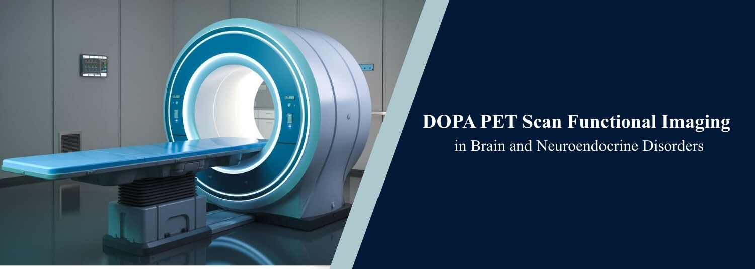
At Molecular Diagnostics and Therapy, we perform DOPA PET scans using modern PET-CT systems to ensure high accuracy and clear imaging.
What Is a DOPA PET Scan?
A DOPA PET scan is a type of Positron Emission Tomography (PET) scan that uses a special radioactive substance called Fluorodopa (FDOPA). This tracer acts in the same manner as L-DOPA, which is used in the formation of dopamine in the body.
Dopamine is an important chemical messenger in the brain. It helps manage movement, coordination, and mood. Neurological problems can occur when dopamine levels are low or abnormal. The DOPA PET scan helps doctors assess dopamine activity in the brain and detect changes not visible on normal MRI or CT scans.
How Does the Scan Work?
The process is simple and painless. A low dose of FDOPA tracer is injected into a vein. The tracer travels through the bloodstream and collects in regions where there is dopamine metabolism or tumor activity. The PET scanner detects signals emitted by the tracer and generates detailed functional images.
Unlike CT or MRI scans, which show structure and shape, DOPA PET identifies how cells are functioning. This makes it especially useful for early diagnosis.
Importance in Brain Disorders
DOPA PET plays a major role in diagnosing Parkinson’s disease. In Parkinson’s, dopamine-producing cells gradually reduce. DOPA PET can detect low dopamine levels even before severe symptoms appear. It also helps doctors differentiate between Parkinson’s disease and other movement disorders.
The scan is also useful in assessing brain tumors such as Glioma. It helps determine whether a tumor has recurred after treatment or if changes seen on MRI are simply scar tissue. This information is crucial for further therapy planning.
Role in Neuroendocrine Disorders
DOPA PET is commonly used to detect certain hormone-producing tumors known as neuroendocrine tumors (NETs), including:
- Neuroendocrine tumors (NETs)
- Pheochromocytoma
- Paraganglioma
These tumors may not always absorb glucose strongly and might not appear clearly on conventional PET scans. DOPA PET is more effective in detecting amino acid and dopamine-related tumor metabolism in such cases.
DOPA PET vs Conventional PET
| Feature | DOPA PET Scan | Conventional PET Scan |
|---|---|---|
| Tracer Used | FDOPA | FDG |
| Detects | Dopamine & Amino Acid Activity | Glucose Metabolism |
| Best For | Parkinson’s & NETs | Most Common Cancers |
| Brain Imaging | More Specific | General Evaluation |
| NET Detection | Highly Sensitive | May Miss Some Cases |
Benefits of DOPA PET
DOPA PET assists in early disease diagnosis, provides functional information, improves diagnostic accuracy, and supports treatment planning. Because it detects changes at the cellular level, it can identify issues before structural damage occurs.
Preparation and Safety
Preparation is usually simple. Patients may be asked to fast for a few hours before the scan. Inform your doctor about any medications, pregnancy, or breastfeeding.
The scan typically takes two to three hours, including waiting time after tracer injection. The radiation dose is controlled and considered safe for medical imaging. Most patients tolerate the procedure well with rare side effects.
When Is DOPA PET Recommended?
Doctors may recommend a DOPA PET scan for symptoms of Parkinson’s disease, unclear brain imaging findings, suspected neuroendocrine tumors, or evaluation of tumor recurrence. The final decision is made by a neurologist or oncologist based on clinical findings.
Conclusion
The DOPA PET scan is an advanced imaging technique that focuses on functional activity rather than structure alone. It plays a crucial role in diagnosing Parkinson’s disease and detecting specific neuroendocrine tumors. By providing early and accurate information, it helps doctors make better treatment decisions and improves patient outcomes.
At Molecular Diagnostics and Therapy, advanced DOPA PET imaging is performed using modern technology and expert supervision to ensure precise diagnosis and reliable results.
Frequently Asked Questions
In News
Get Our Mobile App
for Easy Access
Book tests, view reports, and manage your health records on the go. Experience convenient healthcare with Molecular Diagnostics and Therapy.
- ✔ Book tests & home collection
- ✔ View reports instantly
- ✔ Track health history
- ✔ Get notifications & reminders
- ✔ Easy appointment management
