
Gamma Camera Imaging: How It Helps Diagnose Bone, Kidney, and Thyroid Disorders
Talk to Health Expert

Dr. Nikunj Jain
Co-Founder and HOD - Nuclear Medicine ,MBBS, DRM, DNB, FEBNM, FANMB, Dip. CBNC
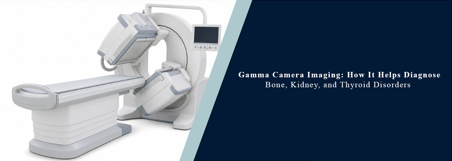
Medical imaging has been revolutionized in modern times, changing the way doctors diagnose disease. Although X-rays, CT scans, and MRI scans give very good pictures of the structure, they do not always reflect organ functioning. This is where gamma scan technology comes into its own. Doctors can use a gamma camera to determine the working health of an organ and also be able to diagnose disease in an early stage.
In a nuclear medicine scan, one injects a radioactive tracer agent in small quantities, which goes to a particular organ. A gamma camera then takes pictures representing the activity of that organ. Since it measures organ function in addition to structure, a nuclear medicine test can be used to find abnormal function before other imaging tests show any abnormality.
Modern gamma camera systems, advanced technology, and the expertise of the physicians and technicians at Molecular Diagnostics and Therapy offer precise and dependable nuclear medicine diagnostic imaging for diverse medical conditions.
What Is Gamma Camera Imaging?
A gamma scan is the name given to a special type of imaging that is used in the field of nuclear medicine. A small dose of radioactive tracer is injected into the body, swallowed, or, rarely, inhaled during the exam. Depending on what the examination is, the tracer accumulates in a certain organ.
The gamma camera captures the tracer's radiation and generates images to reveal the organ's activity. An NM scan can provide helpful data regarding the metabolic activity and well-being of tissues, as opposed to standard imaging.
Why Is Gamma Camera Imaging Important?
A lot of diseases start with non-structural, yet functional, problems. Any functional abnormalities are detectable in the gamma imaging even at an early stage, and doctors can diagnose earlier and start treating the patient appropriately. The technique has widespread applications in cancers, cardiology, nephrology, endocrinology and orthopaedics, in which its value lies in providing additional information not obtained from other imaging techniques.
Bone Scan Test
Bone scan test is one of the most commonly performed tests in nuclear medicine. It can be helpful to identify abnormal bone activity due to fractures, bone infections, arthritis, bone tumors, the complications of osteoporosis, and cancer that has spread to the bones. A bone scan is usually able to detect abnormalities earlier than regular X-rays do.
A bone scan can be used to check for suspicious stress fractures, as well as patients suffering from bone pain that cannot be explained, and those with known cancers that could implant in bone, or long-lasting joint pain. The scan is a measure of bone metabolism, so that often a disease is detected before permanent damage is built up.
Renal Scan Test
A kidney scan test (also called a kidney nuclear scan) measures kidney function instead of just kidney anatomy. This scan assesses the kidneys' ability to filter blood, to make urine, and their drainage into the urinary bladder. It can also measure the blood flow to the kidneys and detect urinary tract blockages.
A renal scan is used in the clinic commonly when a patient has had many urinary infections, kidney blockage, congenital defects in the kidneys, high blood pressure caused by renal disease, or low renal function. The renal scan test is also beneficial for kidney function before and following kidney surgery or kidney transplant.
Thyroid Scan
A thyroid scan is used to determine the activity, position, shape, and size of the thyroid gland. A thyroid scan differs from routine blood tests, which indicate levels of the thyroid hormones, in that it reveals the activity of various sections of the thyroid. The test can be used to determine if the thyroid tissue or glands are overactive, underactive, or if nodules are abnormal.
A doctor might use a thyroid scan to diagnose thyroid cancer or other thyroid problems if a patient has concerns about thyroid function following an abnormality on ultrasound, has hyperthyroidism, an unexplained thyroid hormone disorder, or an enlargement of the thyroid gland. The scan also helps to plan treatment with radioactive iodine and track thyroid problems over the years.
Other Conditions Diagnosed Using Nuclear Medicine
A nuclear medicine scan is used for many medical conditions, though some are more common, such as imaging bones, kidneys, and thyroids. It is also used for much of the time to test heart function, detect infections, search for gastrointestinal bleeding, look up lung diseases, diagnose neurological diseases, and many kinds of cancer. Nuclear medicine imaging plays a significant role in the diagnosis and staging of cancer; in the planning of treatment for cancers and in assessing response to treatment.
How Is a Gamma Scan Performed?
The specific practice is dependent on which organ is being inspected. Once the radioactive tracer is given to the patient, he/she generally has to wait several minutes to hours for the tracer to build up in the tissue of the organ under investigation.
Then the patient is placed on an examination table in a comfortable position, and the gamma camera is taken around the body to acquire a series of detailed images. There is no pain associated with the underlying scan, and it normally can be completed in 20 - 60 minutes, depending on the type of scan. Patients must be completely motionless during image acquisition to obtain good and clear images to reduce errors.
Is Gamma Camera Imaging Safe?
The amount of radioactivity required for a gamma scan is extremely low and is controlled to minimise exposure. Medical radiation very rarely poses a risk compared to the great benefit obtained from the final diagnosis. There are hardly any cases of serious allergic reactions to the radioactive tracer. If a woman is pregnant or breastfeeding, she should tell her physician before having a nuclear medicine examination; a different type of imaging sometimes may be suggested.
Understanding the Scan Report
A radiotracer is then given to the patient, and a nuclear medicine physician thoroughly interprets the images and issues a comprehensive report. The report includes a description of how the organ is functioning, the presence of a possible normal or abnormal uptake of tracer, and evidence of the presence of disease or impairment of organ function.
These findings, combined with the patient's symptoms, medical history, laboratory results, and other imaging data, will help the doctor make an accurate diagnosis and decide which treatment is best.
Choosing the Right Nuclear Medicine Centre
The accuracy of a nuclear medicine scan depends not only on advanced imaging equipment but also on experienced specialists who interpret the results correctly. If you are searching for a nuclear medicine centre near me, choose a facility equipped with modern gamma camera technology, qualified nuclear medicine physicians, and high-quality reporting standards.
At Molecular Diagnostics and Therapy, patients receive comprehensive nuclear medicine services, including bone scan test, renal scan test, thyroid scan, and other specialised gamma camera imaging studies performed using advanced technology and expert clinical interpretation.
Conclusion
Diagnostic medicine has come a long way since the days of X-rays. Nowadays, doctors can use a gamma camera to assess organ function even when structural disease is not yet apparent. This technology is used to diagnose and develop treatment strategies at an early stage and monitor disease, and is used in bone scan tests, renal scan tests, thyroid scans, and other nuclear medicine-based examinations.
Our modern imaging systems, experienced specialists, and sound reporting at Molecular Diagnostics and Therapy set the benchmark for high-quality nuclear medicine services, bringing the utmost level of diagnosis and timely diagnosis to their patients.
Frequently Asked Questions
In News
Get Our Mobile App
for Easy Access
Book tests, view reports, and manage your health records on the go. Experience convenient healthcare with Molecular Diagnostics and Therapy.
- ✔ Book tests & home collection
- ✔ View reports instantly
- ✔ Track health history
- ✔ Get notifications & reminders
- ✔ Easy appointment management
