
PET-CT Scan: Purpose and Procedure
Talk to Health Expert

Dr. Nikunj Jain
Co-Founder and HOD - Nuclear Medicine ,MBBS, DRM, DNB, FEBNM, FANMB, Dip. CBNC
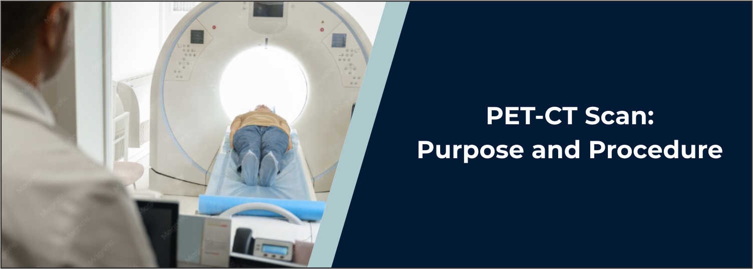
A PET-CT scan is an advanced imaging test that integrates two
types of imaging, positron emission tomography (PET) and computed tomography
(CT) into one exam. It offers data on the function of tissues and organs and
detailed images showing the structure of tissues and organs. PET-CT is
frequently used to help determine if cancer has spread, to help plan treatment,
to determine the response to treatment, and to look for signs of cancer
returning. Additionally, it can be used for certain heart and neurological
diseases.
PET-CT scans are done at Molecular Diagnostics and Therapy to
facilitate the investigation and management of various health problems as
indicated by a physician. This blog describes the PET-CT scan procedure, the
preparation for PET-CT, the duration of PET-CT, and other important pet ct test
information.
What Is a PET-CT Scan?
PET-CT is an integrated imaging study that combines the
information of two types of imaging tests. The PET component involves a
radioactive material: a radiotracer. A tracer that is commonly used is
fluorodeoxyglucose (FDG), a molecule that is structurally similar to glucose
and is used in the body. Regions where the metabolism is more intense may
absorb more FDG and may become more prominent on PET images.
The CT component takes X-rays of the body to make detailed
pictures of the body. Together with the PET picture, the doctor can also see
where a part of the body is abnormal, along with how active that area is. This
combination can give you information that might be unable to be acquired with
either alone.
Why Is a PET-CT Scan Done?
Doctors may recommend a
PET-CT scan for several reasons, particularly during cancer evaluation and
treatment. Common uses include:
- Detecting or evaluating certain cancers
- Determining
whether cancer has spread to other parts of the body
- Assessing
response to cancer treatment
- Checking
for possible recurrence after treatment
- Helping
with treatment planning
- Evaluating
selected neurological conditions
- Assessing
certain heart-related conditions
The exact reason for a
PET-CT depends on the patient's symptoms, diagnosis, previous test results, and
treatment history.
PET-CT Scan Preparation
Because of the effects that food consumption and blood glucose
levels have on the distribution of FDG, proper PET-CT scan preparation is
important. In several FDG PET-CT studies, patients are asked to avoid food for
a few hours before the study. To help maintain hydration, which usually
includes drinking plain water is usually allowed and encouraged, though drinks
high in sugar or calories should be avoided unless directed otherwise by the
medical team.
Individuals with diabetes may be given special instructions for
preparation as blood glucose levels can affect the acquisition of good images
during an FDG PET-CT examination. Before the scan, tell the medical team if
you:
- Have diabetes
- Are
pregnant or think you may be pregnant
- Are
breastfeeding
- Take
regular medicines
- Have
any allergies
- Have
recently had an infection, surgery, or other significant medical condition
You should follow the
preparation instructions provided by the imaging centre because requirements
can vary depending on the type of PET-CT being performed.
What Happens During the PET-CT Scan Procedure?
The PET-CT scanning process normally starts with a technologist
verifying your information and preparing you for the test. A tiny IV is
typically inserted into an arm vein. The prescribed radiotracer is then
injected through the IV. Following the injection, it is important to be very
still as the radiotracer moves around your body.
This is usually about 30-60 minutes for FDG PET-CT. At this time,
you might be asked to keep still and not move about or talk. After uptake is
done, you are positioned on the scanning table. Slow motion: The table is
slowly moved into the PET-CT scanner to get the image. The purpose of the scan
is to obtain clear images, and this requires the subject to remain still during
the scan.
PET-CT Scan Duration
A PET-CT scan lasts for two parts: (1) preparation and tracer
injection (2) the scan. Typically, it takes 30 to 60 minutes for the
radiotracer to get distributed around the body. The actual scanning time is
typically based on the area of the body being scanned and can vary from 30
minutes for the most common type of scan for most patients. So, as a general
rule, patients can expect to be at the centre well beyond the time it takes to
be scanned. Examination may be longer because of additional imaging, certain
imaging procedures, or contrast or special protocols.
What Does a PET-CT Scan Feel Like?
Typically, PET-CT is painless except for the slight discomfort
associated with inserting its IV line. The IV might cause a little stinging
when it is inserted. In some people, there may be a brief cold sensation in the
area of the injection, or the needle may feel a bit cool. When taking a scan,
you must remain still on the examination table. It may cause slight discomfort
for those who must not move in one position for too long. The opening in the
scanner is large and in the shape of a doughnut. People don't spend a long time
inside a long, narrow tube as they do with an MRI scanner.
PET-CT Scan Test Details
The exact PET-CT scan
test details depend on the reason for the examination and the radiotracer being
used. A typical FDG PET-CT may involve:
1. Patient registration
and preparation:
Your medical history, medicines, diabetes
status, and other relevant information may be reviewed.
2. Blood glucose
assessment:
For FDG PET-CT, blood glucose may be checked
before the tracer is administered because abnormal glucose levels can affect
the examination.
3. Radiotracer
injection:
A small amount of the prescribed radiotracer is
injected through an IV.
4. Uptake period:
You rest quietly while the tracer distributes
through the body.
5. CT scan:
The CT component provides detailed anatomical
information.
6. PET scan:
The PET component records the distribution of
the radiotracer and provides information about metabolic activity.
7. Image interpretation:
The resulting images are reviewed by
appropriately trained imaging specialists, and a report is prepared for the
referring doctor.
What Should You Do After a PET-CT Scan?
After the examination,
most people can return to their normal activities unless their doctor or
imaging team provides different instructions. Drinking plenty of water is
generally encouraged after many PET examinations because the radiotracer is
gradually eliminated from the body, including through urine. If you are
breastfeeding, pregnant, or have another condition requiring special
precautions, follow the specific instructions provided by your healthcare team.
Is PET-CT Safe?
PET-CT involves exposure
to radiation from both the radioactive tracer used for PET and the CT
component. The amount is carefully considered according to the clinical need
for the examination. The radiotracer used for PET has a short physical
half-life, and its radioactivity decreases naturally over time. Much of the
tracer is also eliminated from the body through urine or stool. Before the
examination, inform your doctor or imaging team if you are pregnant or
breastfeeding so that the benefits and appropriate precautions can be
considered.
What Can a PET-CT Scan Show?
A PET-CT scan can
provide information about both the structure and metabolic activity of tissues.
In cancer care, areas showing increased tracer uptake may indicate increased
metabolic activity. However, increased uptake does not always mean cancer
because inflammation and infection can also cause increased uptake.
Therefore, PET-CT findings are interpreted together with symptoms, medical history, physical
examination, laboratory tests, biopsy results, and other imaging findings when
required. A PET-CT scan is an important diagnostic and monitoring tool, but its
findings should always be interpreted in the appropriate clinical context.
PET-CT at Molecular Diagnostics and Therapy
At Molecular Diagnostics
and Therapy, PET-CT services are available for patients who require advanced
nuclear medicine imaging based on their doctor's recommendation. The
examination can provide combined metabolic and anatomical information that may
help doctors evaluate disease, assess its extent, monitor treatment response,
or look for recurrence when clinically appropriate. Patients should follow the
centre's specific preparation instructions before arriving for their
appointment to help ensure that the examination can be performed properly.
Conclusion
In a PET–CT scan, the patient is given a combination of PET scan
and CT scan, which gives information about both the metabolic activity and the
anatomical structure of tissues and organs. PET-CT is used in the evaluation,
staging, treatment assessment, and surveillance of selected cancers. The
correct preparation procedures, especially in the case of PET-CT imaging with a
PET radiotracer, are crucial. Patients must also withhold food for a few hours,
refrain from sugary beverages, and drink plenty of plain water. Medication
instructions may vary, particularly for patients with diabetes.
In general, the PET-CT scanning process is carried out through the
injection of a radio-tracer into the bloodstream via intravenous drip, a brief
rest time for uptake, followed by PET and CT imaging. The examination itself
takes about 30 minutes to give the PET scan; the actual scanning phase is
usually about 30 minutes, though the length of time after tracer uptake varies
depending on the examination. For Molecular Diagnostics and Therapy, PET-CT
imaging is used to aid in the assessment and management of various illnesses,
as indicated by clinical conditions. Best results are achieved when patients
follow all the preparation instructions given before examination.
Frequently Asked Questions
In News
Get Our Mobile App
for Easy Access
Book tests, view reports, and manage your health records on the go. Experience convenient healthcare with Molecular Diagnostics and Therapy.
- ✔ Book tests & home collection
- ✔ View reports instantly
- ✔ Track health history
- ✔ Get notifications & reminders
- ✔ Easy appointment management
