
What Is a PET-CT Scan?
Talk to Health Expert

Dr. Nikunj Jain
Co-Founder and HOD - Nuclear Medicine ,MBBS, DRM, DNB, FEBNM, FANMB, Dip. CBNC
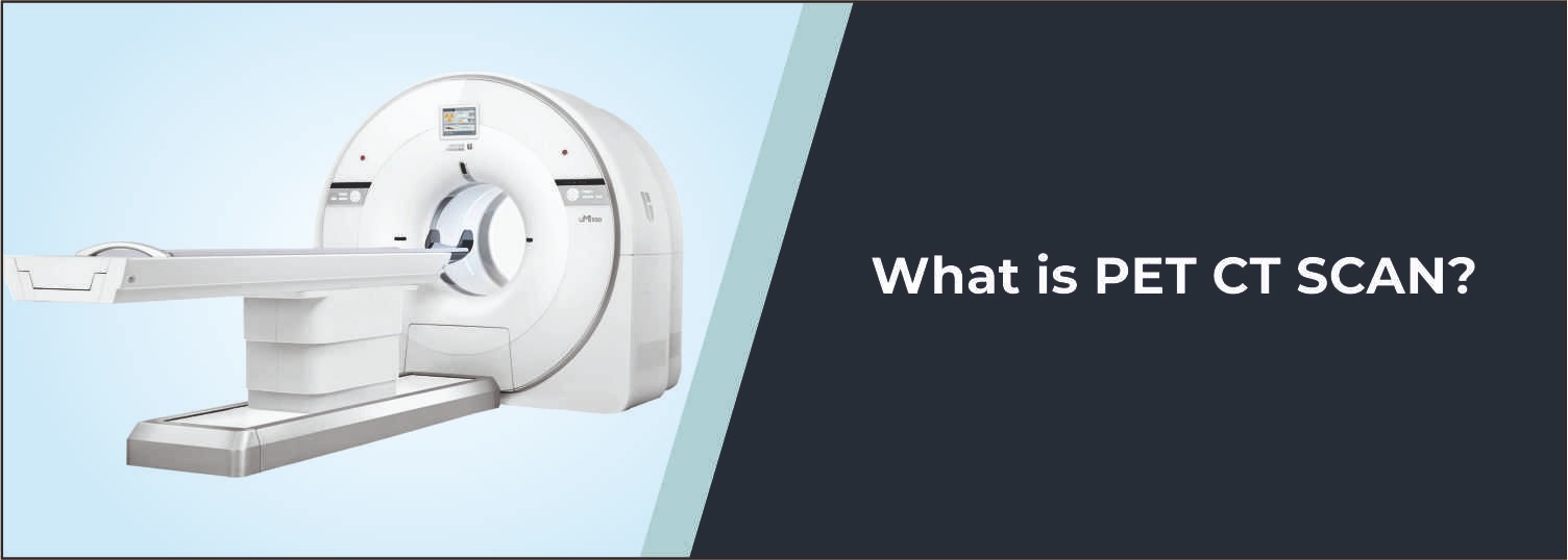
PET-CT imaging is a modern procedure
that combines two imaging technologies into one: Positron Emission Tomography
(PET) and Computed Tomography (CT). PET shows activity and metabolism in
tissues, while CT provides detailed images of the body's internal structures.
The combination of these two types of information is used to help a physician
look not only at the location of abnormal areas but also at their activity
level.
PET-CT is widely used in cancer
evaluation and management, especially for some cancers, to determine if the
cancer has metastasized (spread), to plan cancer treatment, and to look for
signs of response to cancer treatment. Also, it is used in specific heart and
neurological diseases as appropriate in individual cases.
What Is a PET-CT Scan?
A PET-CT scan is a PET scan performed
under the same conditions as those of a computed tomography (CT) scan, which
also uses a computed tomography scanner. A tiny dose of radioactive material,
called a radiotracer, is administered before the scan, typically intravenously.
Many PET scans use a glucose analogue labeled with fluorine-18, known as FDG
(fluorodeoxyglucose). Areas that are undergoing metabolic activities different
from those of normal tissues appear differently on PET images.
However, the uptake rate does not only
have to do with cancer, as it occurs in some normal tissues or inflammatory
diseases as well. The CT gives information about the size, shape, and location
of organs and other structures. PET-CT information is used together to relate
metabolic activity with anatomical information.
Why Is PET-CT Done?
A PET-CT is recommended for several
reasons, primarily to examine cancer. It aids in the diagnosis or evaluation of
types of cancer, and is helpful in determining if a cancer has spread to other
areas of the body and in providing information that could be beneficial in
planning treatment. PET-CT is used after, or during treatment, to see how the
disease is responding and, in rare cases, to detect signs of recurrence.
Whether PET-CT has a primary or secondary role is dependent on the kind of
disease, the clinical question asked, prior tests performed, and the therapy
planned.
How Does PET-CT Work?
PET and CT scans both give different
information. The PET scan examines biological or metabolic activity. A
radiotracer enters the bloodstream and is taken up by tissues. The PET scanner
records the signals emitted by the radiotracer and generates images of where
the tracer is located in the body. A CT scan produces detailed cross-sectional
images of the structures in the body using X-rays. The images are fused back
together, and the doctor compares the region of the increased or decreased
uptake with the corresponding anatomical region. This gives data that can be
more beneficial than that obtained from either kind of imaging alone.
PET-CT Scan Procedure
Typically, the PET-CT procedure starts
with preparation before injection of the radiotracer. The patient should not
consume food for a few hours before the PET-CT and is instructed to drink
plenty of water before the scan as appropriate for the type of PET-CT and
radiotracer. An IV line is typically inserted into a vein, and the radiotracer
is injected by a health care professional. The patient then lies still so that
the tracer passes through the body, concentrating in the tissues that are being
examined. This is a typical time of waiting of 30-60 minutes.
The patient then rests on a table that
is used to conduct the scan. The table runs in the PET-CT scanner, and the
images are taken. During the scan, it is very important not to be in movement,
to avoid the image being affected. The scanning time is usually approximately
30 minutes, although this can be longer for some imaging procedures, and it can
depend on the examination.
How Should You
Prepare for a PET-CT Scan?
Depending on the type of PET-CT, one
may need to prepare for the CT. Patients are usually advised to not eat for a
certain time before an FDG PET-CT exam and will be able to drink water during
the procedure. Blood glucose might be especially critical for FDG PET-CT as
fluctuations in blood glucose or insulin levels may impact the distribution of
the tracer and assessment results of the examination.
If they have been diagnosed with
diabetes, have been prescribed any medication, have had any recent illness, may
have had any type of allergy, or have been pregnant or breast feeding should be
conveyed to the medical team before examination. Specific preparations are
explained by the imaging centre according to the procedure of the examination.
What Are the Benefits
of PET-CT?
One very important advantage of PET-CT
is that the functional information that is provided by PET can be overlaid onto
the anatomical information provided by CT. This can aid in identifying abnormal
metabolic rate locations and, indeed, their position in the body. PET-CT can be
used to evaluate the disease, stage the cancer, plan treatment, and assess
response to treatment in cancer care. One added advantage is that PET and CT
exams can be performed at the same time, and the two types of data can be
correlated without having to book a separate appointment for the two scans.
Is PET-CT Only Used
for Cancer?
No, PET-CT is used in oncology but has
other uses in medicine. Depending on the radiotracer and clinical need, PET
imaging can be used to assess some heart diseases and neurological disorders.
As a case in point, PET can be used to evaluate metabolism and function in the
brain or heart, depending on the radiotracer and clinical indication. The type
of radiotracer and imaging procedure depend on the condition that is being
studied.
Is PET-CT Safe?
PET-CT involves exposure to radiation because it uses a radioactive
tracer and CT X-rays. The radiotracer used for PET is given in a small amount
and loses its radioactivity over time. The route of elimination depends on the
radiotracer. As with other medical imaging procedures, doctors consider the
expected clinical benefit before recommending the examination. Patients should
inform the medical team if they are pregnant or breastfeeding because special
precautions may be required.
What Does a PET-CT
Report Show?
A PET-CT report describes findings seen on the PET and CT images.
Depending on the examination, the report may discuss areas of increased or
decreased tracer uptake, anatomical abnormalities, and other relevant findings.
The report is interpreted by a suitably trained specialist and should be
considered together with the patient's symptoms, medical history, previous
investigations, and other clinical information.
Importantly, an area of increased tracer uptake does not automatically
mean cancer. Some infections, inflammation, and normal tissues can also show
increased uptake. Therefore, PET-CT findings may need to be correlated with
other tests or clinical information.
What Happens After a
PET-CT Scan?
After the scan, most patients can return to their normal activities
unless their healthcare provider gives different instructions. Drinking
adequate water may help the body eliminate the radiotracer through urine. The
radiotracer loses radioactivity naturally over time, and the healthcare team
may provide specific instructions depending on the radiotracer and examination.
Limitations of PET-CT
PET-CT is a valuable diagnostic tool, but it does not provide all the
information needed in every medical situation. PET images generally have lower
anatomical resolution than CT or MRI, although PET provides functional
information that these imaging techniques may not provide in the same way. In
addition, blood glucose levels can affect some PET examinations, and very small
or less metabolically active abnormalities may not always be clearly detected.
For this reason, doctors may recommend other imaging tests, laboratory tests,
or a biopsy depending on the suspected condition.
PET-CT at Molecular
Diagnostics and Therapy
At Molecular Diagnostics and Therapy, advanced diagnostic imaging
services include Digital PET-CT, which can provide both functional and
anatomical information in a single examination. The appropriate PET-CT protocol
and radiotracer depend on the patient's medical condition and the clinical
question. Our diagnostic team follows the required preparation and imaging
protocols to support doctors in evaluating disease and planning further
management.
Conclusion
PET-CT is a powerful study which
integrates the functional information provided by the PET with the detailed
anatomical images obtained with the CT. PET-CT is widely used in cancer
evaluation for disease assessment, staging, treatment planning, and assessment
of treatment response where clinically appropriate
The PET-CT process includes giving the patient the radiotracer, then a waiting time and imaging. However, good preparation is necessary, particularly when taking an examination with FDG, and patients must follow the instructions of their healthcare team. Although very useful, PET-CT is not a definitive single test to diagnose all conditions. Doctors use the information from PET scans and other tests along with symptoms, medical history, and, if needed, tissue diagnosis to determine the patient's disease.
Frequently Asked Questions
In News
Get Our Mobile App
for Easy Access
Book tests, view reports, and manage your health records on the go. Experience convenient healthcare with Molecular Diagnostics and Therapy.
- ✔ Book tests & home collection
- ✔ View reports instantly
- ✔ Track health history
- ✔ Get notifications & reminders
- ✔ Easy appointment management
