
DOTA PET Scan vs Conventional PET Scan Key Differences Explained
Talk to Health Expert

Dr. Nikunj Jain
Co-Founder and HOD - Nuclear Medicine ,MBBS, DRM, DNB, FEBNM, FANMB, Dip. CBNC
In recent years, there has been a lot of development in nuclear medicine, which has significantly helped and improved the way doctors detect and manage cancer. And one of such important innovations is PET scans, as they provide detailed and functional imaging of the body. But all PET scans are not the same. Two of the most commonly used PET scans are the DOTA PET Scan and the Conventional PET Scan (FDG PET-CT). These both type of scans use radioactive tracers which detect diseases. But their applications, accuracy, and importance clinically are very different.
At Molecular Diagnostics and Therapy, we use PET imaging technology, which is advanced and used to provide a detailed cancer diagnosis with treatment planning according to the patient.
Understanding Conventional PET Scan
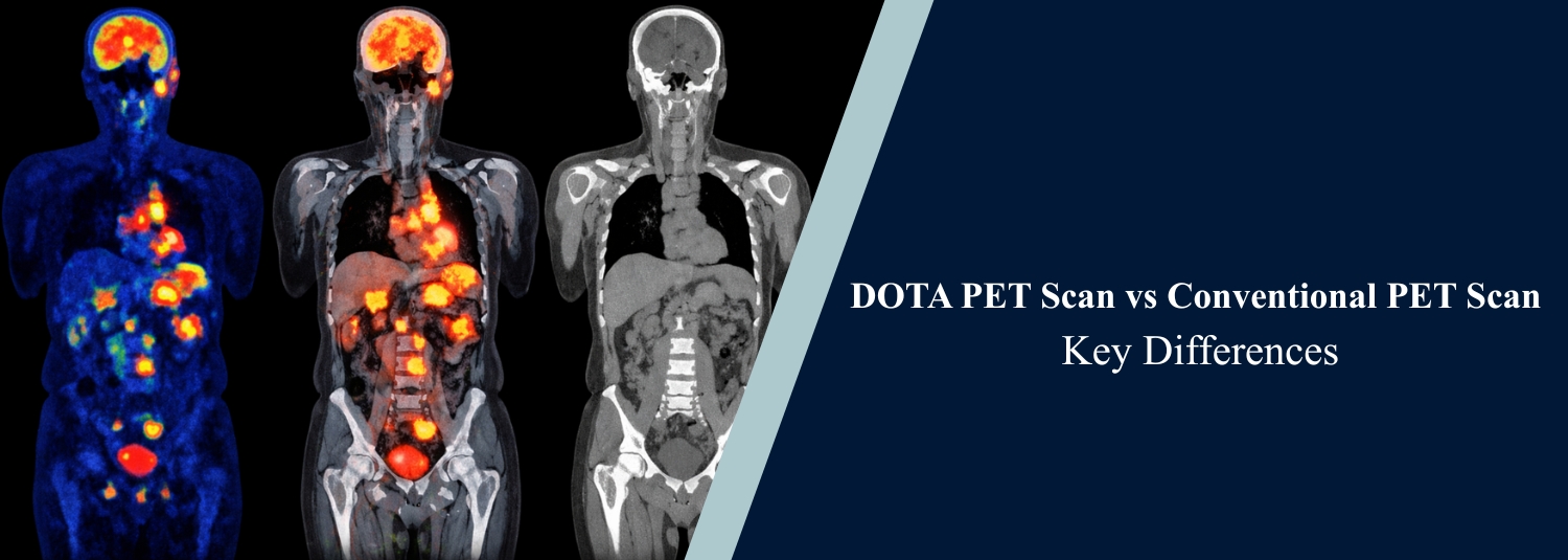
The standard PET image is also referred to as the FDG PET-CT. It involves the usage of a radioactive glucose known as fluorodeoxyglucose (FDG). Cancer cells tend to consume greater glucose levels than normal cells, and hence, regions that show more metabolic activity light up the scan.
In the treatment of cancers, this scan is mostly used as it helps detect many common types of cancer, including lung, breast, lymphoma, colon, and head and neck tumors. It is also very helpful for such tasks as staging disease, detecting metastasis, and evaluating how well a patient is responding to chemotherapy or radiation therapy.
Conventional PET focuses primarily on metabolic activity, meaning it highlights tissues that are using more energy than normal.
Understanding DOTA PET Scan
The scan is the DOTA PET, which is designed to locate neuroendocrine tumors (NETs). It does not examine the metabolism of glucose but rather employs a tracer, which attaches to somatostatin receptors that the NET cells possess. The steadily growing number of NETs can be challenging to see with the conventional PET scan since many of them grow slowly and may not consume a lot of glucose. Due to receptors, DOTA PET observes the tumors, thus it is extremely effective when it comes to locating NETs, even when they are small or when they are at an early stage.
The targeted method provides a more precise insight into small or early NETs and assists in directing the treatment, such as peptide receptor radionuclide therapy (PRRT).
Key Differences at a Glance
Below is a simplified comparison to better understand how these two scans differ:
When Do Doctors Recommend Conventional PET?
The doctors tend to recommend an FDG PET scan in case they suspect that there is a normal solid tumor or lymphoma. It is particularly helpful where the cancer is aggressive and consumes a lot of glucose. It is also preferred for:
Evaluating suspected cancer spread
Checking for recurrence after treatment
Monitoring chemotherapy effectiveness
Investigating unexplained weight loss or abnormal tumor markers
Since it applies to a wide range of cancers, FDG PET is the most conventional PET scan performed in the world.
When Is DOTA PET Recommended?
A DOTA PET scan has been suggested by doctors when the existence of a neuroendocrine tumor is determined. They can recommend it in case blood tests indicate a NET or when previous imaging failed to provide a definite result. It is especially helpful for:
Detecting small, slow-growing NETs
Locating unknown primary NET tumors
Evaluating disease spread in NET patients
Planning targeted radionuclide therapy
In the case of NETs, DOTA PET often provides a lot of more detailed information compared to a standard PET.
Safety and Procedure
The scans are both conducted similarly. A radioactive tracer is injected in a small amount into a vein. Images are captured by the scanner after a while. The entire process is not time-consuming, taking just a few hours. Both tests are safe. The dose of radiation is also well-thought-out and does not exceed the medical safety limits. The tracer exits the body prematurely.
We have strong safety regulations and a high-performance imaging system at Molecular Diagnostics and Therapy, whereby we ensure that the patients are comfortable and the results are accurate.
Which One Is Better?
It is not a question of which scan is better, but which scan is appropriate in this case. PET Conventional PEPT is ideal with most common cancers, whereas DOTA PET is by far superior in neuroendocrine tumors. Your oncologist or nuclear medicine specialist will choose the scan based on:
Type of suspected tumor
Clinical history
Blood test results
Previous imaging findings
Treatment planning needs
Choosing the right imaging method improves diagnostic confidence and treatment outcomes.
Conclusion
Both DOTA PET and the conventional PET are major milestones in cancer imaging. Most common cancers are the ones that are studied by FDG PET-CT, while neuroendocrine tumors have been transformed by DOTA PET as a mode of detection and treatment. This understanding of the differences assists the patients in obtaining the most suitable test regarding the condition.
Molecular Diagnostics and Therapy offers PET imaging at the highest possible level to deliver credible, patient-centered care and contribute to the enhancement of cancer management.
Frequently Asked Questions
In News
Get Our Mobile App
for Easy Access
Book tests, view reports, and manage your health records on the go. Experience convenient healthcare with Molecular Diagnostics and Therapy.
- ✔ Book tests & home collection
- ✔ View reports instantly
- ✔ Track health history
- ✔ Get notifications & reminders
- ✔ Easy appointment management
