
Bone Scintigraphy – Purpose, Procedure & Risks
Talk to Health Expert

Dr. Nikunj Jain
Co-Founder and HOD - Nuclear Medicine ,MBBS, DRM, DNB, FEBNM, FANMB, Dip. CBNC
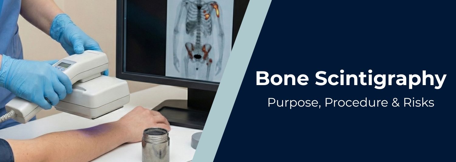
Bone scintigraphy, also known as a bone scan, is a nuclear medicine imaging technique used to evaluate abnormalities in the bones. It involves the injection of a small amount of radioactive tracer (usually Technetium-99m) into a vein, which is absorbed by bone tissue and detected using a gamma camera.
Areas with abnormal tracer uptake (increased or decreased) can indicate a variety of bone conditions. This scan is particularly useful in detecting bone infections, cancer spread, hidden fractures, or metabolic bone diseases that might not be visible on standard X-rays or CT scans.
Because of its high sensitivity, bone scintigraphy can detect issues earlier than many other imaging methods. In a diagnostic setting like Delhi, where advanced nuclear medicine services are available, bone scintigraphy is routinely used by orthopedic surgeons, oncologists, and rheumatologists for accurate diagnosis and treatment planning.
Purpose of Bone Scintigraphy
Bone scintigraphy helps visualize physiological activity in bones, not just anatomical structure, making it extremely valuable in diagnosing or monitoring bone disorders.
Major Clinical Purposes Include:
-
Detection of Bone Metastases
Used to check if cancers (e.g., breast, prostate, or lung) have spread to the bones. -
Diagnosing Bone Infections (Osteomyelitis)
Helps detect infections early and differentiate infected areas from healthy bone tissue. -
Identifying Stress Fractures
Highly sensitive in spotting stress or micro-fractures, especially in athletes and the elderly. -
Evaluation of Bone Pain
Useful when the cause of persistent or vague bone pain is unclear. -
Detection of Arthritis and Joint Inflammation
Reveals active inflammation in joints—especially beneficial in diagnosing rheumatoid arthritis or similar inflammatory conditions. -
Assessing Bone Tumors
Differentiates between benign and malignant bone lesions; also monitors tumor response to treatment. -
Investigation of Unexplained Fractures or Prosthesis Issues
Detects loosening or complications with orthopedic implants. -
Evaluating Paget’s Disease of Bone
Monitors areas of abnormal bone remodeling.
Bone scintigraphy is often performed as a whole-body scan, especially when evaluating cancer spread or systemic bone disease.
Procedure: What to Expect During a Bone Scan
Bone scintigraphy is typically conducted in two parts on the same day:
one for tracer injection and one for imaging, spanning about 3–4 hours.
1. Before the Scan
-
Usually, no fasting or special preparation is needed.
-
Inform your doctor if you are pregnant, breastfeeding, or recently had imaging with contrast.
-
Remove any metallic jewelry or accessories.
2. Tracer Injection
-
A small amount of radioactive tracer (Technetium-99m) is injected into a vein in your arm.
-
The tracer travels through the bloodstream and accumulates in bones with high metabolic activity.
-
The injection is safe and typically causes no side effects.
3. Waiting Period (2–3 Hours)
-
Wait 2–3 hours for the tracer to distribute and absorb into your bones.
-
You may be encouraged to drink fluids and empty your bladder frequently to improve scan quality.
4. Image Acquisition
-
You’ll lie on a scanning table while a gamma camera slowly moves over your body.
-
The camera captures images of areas with abnormal tracer concentration.
-
Remaining still during imaging is crucial for accurate results.
5. After the Scan
-
Resume normal activities immediately.
-
Drink plenty of water to flush the tracer from your body.
-
A nuclear medicine specialist will analyze the scan, and your doctor will receive the report in 24–48 hours.
Risks of Bone Scintigraphy
Bone scans are generally safe and well-tolerated, with minimal risks involved.
1. Radiation Exposure
-
The radiation dose is low and comparable to a few X-rays.
-
Not advised during pregnancy unless absolutely necessary.
2. Allergic Reactions
-
Rare and usually mild (e.g., itching, rash).
3. Discomfort from Injection
-
Minor pain, swelling, or bruising at the IV site may occur.
4. Radioactive Material Precautions
-
The tracer loses radioactivity quickly and is excreted via urine.
-
Normal hygiene practices like frequent urination and handwashing are sufficient post-scan.
Overall, the diagnostic benefits far outweigh the minimal risks associated with the scan.
Conclusion
Bone scintigraphy is a powerful, early-detection tool that highlights functional abnormalities in the skeleton before structural changes appear. It is especially valuable in fields like oncology, orthopedics, and rheumatology, where early intervention can significantly improve outcomes.
In Delhi, patients with bone pain, cancer history, or suspected infections have easy access to bone scans at modern nuclear medicine centers. The test is safe, accurate, and essential for personalized treatment planning in complex or ambiguous cases.
In News
Get Our Mobile App
for Easy Access
Book tests, view reports, and manage your health records on the go. Experience convenient healthcare with Molecular Diagnostics and Therapy.
- ✔ Book tests & home collection
- ✔ View reports instantly
- ✔ Track health history
- ✔ Get notifications & reminders
- ✔ Easy appointment management
