
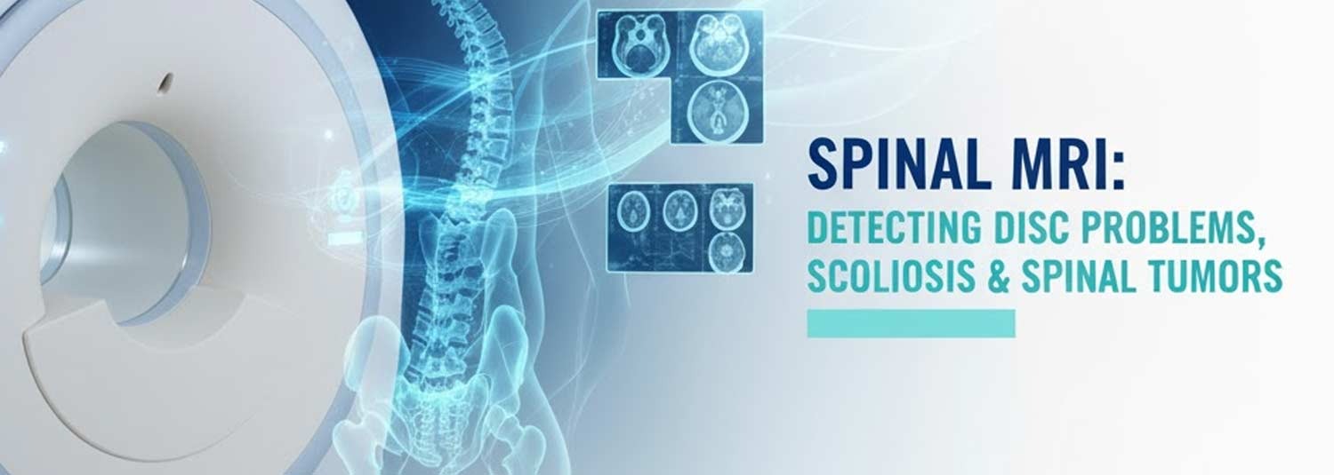
Spinal MRI: Detecting Disc Problems, Scoliosis & Spinal Tumors
Talk to Health Expert

Dr. Nikunj Jain
Co-Founder and HOD - Nuclear Medicine ,MBBS, DRM, DNB, FEBNM, FANMB, Dip. CBNC
Back pain, nerve tingling, or sudden weakness can be frightening. Often, the next step in figuring out what’s happening is imaging. Among the many tests available, a spinal MRI (Magnetic Resonance Imaging) is one of the most advanced and detailed tools doctors use.
But what exactly is a spinal MRI, when is it needed, and what conditions can it detect—like disc problems, scoliosis, and spinal tumors? Let’s break it down in simple, easy-to-understand language.
What Is a Spinal MRI?
A spinal MRI scan is a non-invasive imaging test that uses strong magnetic fields and radio waves to create highly detailed pictures of your spine. Unlike X-rays or CT scans, an MRI does not use radiation, making it a safer option for repeated imaging.
Doctors order a spine MRI scan when they need to look closely at:
- The vertebrae (bones of the spine)
- Intervertebral discs (the cushions between the bones)
- The spinal cord and nerves
- Surrounding tissues, ligaments, and muscles
It’s especially helpful in diagnosing back pain causes that aren’t visible on regular X-rays.
How Is a Spinal MRI Done?
The spinal MRI procedure is simple but requires patience:
- You’ll lie on a sliding table that moves into the MRI machine.
- The machine takes detailed pictures of your spine, sometimes from your neck down to your lower back.
- The test usually takes 30–60 minutes.
- In some cases, you’ll receive contrast dye (usually gadolinium) through an IV to highlight tumors, infections, or nerve issues.
- The machine makes loud tapping or thumping noises, but earplugs or headphones are provided.
For people with claustrophobia, an open MRI for spine scans may be recommended.
Spinal MRI for Disc Problems
One of the most common reasons for getting a spine MRI scan is suspected disc problems, such as:
- Herniated disc (slipped disc): When the soft inner part of a disc bulges out and presses on nerves.
- Degenerative disc disease: Age-related wear and tear that causes back pain.
- Bulging disc: A disc that sticks out but hasn’t fully ruptured.
A spinal MRI gives a clear view of the disc, nerves, and surrounding structures—helping doctors decide whether physical therapy, medication, or surgery is needed.
Spinal MRI for Scoliosis
Scoliosis is a sideways curvature of the spine, often diagnosed in childhood or adolescence. While X-rays are usually the first tool for scoliosis detection, a spinal MRI is sometimes recommended to:
- Check for spinal cord abnormalities.
- Rule out underlying causes such as tumors or cysts.
- Plan surgery in severe cases.
Compared to X-rays, a scoliosis MRI provides much more detail about soft tissues and nerves.
Spinal MRI for Tumors
A spinal cord tumor or other spinal growth is a serious condition that requires early detection. A spine MRI with contrast is the best imaging test for:
- Detecting spinal cord tumors.
- Determining whether cancer has spread to the spine.
- Identifying infections or inflammatory conditions that mimic tumors.
The National Cancer Institute (NCI) recognizes MRI as a critical tool for detecting and monitoring spinal tumors.
When Do You Need a Spine MRI?
Doctors may order a spinal MRI scan if you have:
- Chronic or unexplained back pain.
- Numbness, weakness, or tingling in the arms or legs.
- Symptoms of sciatica (nerve pain shooting down the leg).
- Suspected tumors or infections.
- Abnormal findings on an X-ray or CT scan.
- Signs of nerve compression.
Spinal MRI vs Other Imaging Tests
- X-rays: Good for bones and scoliosis but not soft tissues.
- CT scans: Useful for trauma or bone injuries but involve radiation.
- Spinal MRI: Best for detailed imaging of discs, nerves, tumors, and spinal cord.
Risks and Safety of Spinal MRI
A spine MRI is generally safe, but here are a few things to know:
- No radiation is used.
- Some patients may feel discomfort from lying still.
- Contrast dye risks are rare but can include allergic reactions.
- Patients with metal implants, pacemakers, or cochlear implants should inform their doctor before scheduling.
The American College of Radiology (ACR) has clear safety guidelines to ensure MRIs are safe for most patients.
Key Facts from Trusted Organizations
- American College of Radiology (ACR): MRI is the gold standard for spinal imaging when soft tissue or nerve involvement is suspected.
- National Institute of Arthritis and Musculoskeletal and Skin Diseases (NIAMS): Recommends MRI for diagnosing spinal disc problems and degenerative conditions.
- National Cancer Institute (NCI): MRI scans are critical for early detection and management of spinal tumors.
- World Health Organization (WHO): Identifies spine imaging as essential in managing back pain, one of the most common global health problems.
The Role of Molecular Diagnostics in Spinal Conditions
While a spinal MRI shows images of bones, discs, and nerves, molecular diagnostic testing provides insights at the genetic and biochemical level. Together, they:
- Detect cancers affecting the spine earlier.
- Differentiate between benign and malignant spinal tumors.
- Help doctors create more personalized treatment plans.
- Improve overall accuracy of diagnosis when combined with imaging.
This makes the pairing of spinal MRI + molecular diagnostics the most advanced way to detect and treat spine-related conditions.
Final Thoughts
A spinal MRI is one of the most powerful tools available for detecting disc problems, scoliosis, and spinal tumors. It provides highly detailed images without radiation, making it safer and more effective than many other imaging tests.
If you’re struggling with unexplained back pain, numbness, or neurological symptoms, talk to your doctor about whether a spinal MRI is right for you. And remember—when combined with molecular diagnostic testing, it gives doctors the clearest picture possible for diagnosis and treatment.
Take control of your spinal health today. Early detection leads to better outcomes.
10 FAQs About Spinal MRI
What is a spinal MRI?
A spinal MRI is a scan that creates detailed images of your spine using magnets and radio waves.
Does a spine MRI use radiation?
No, MRIs do not use radiation.
How long does a spinal MRI take?
Usually between 30 and 60 minutes.
Why would a doctor order a spine MRI?
To check for disc problems, scoliosis, tumors, or nerve compression.
What’s the difference between a spine MRI and a CT scan?
MRI gives more detail of soft tissues, while CT is faster and better for bone injuries.
Is a spinal MRI safe?
Yes, it’s safe for most people. Patients with certain implants may not be eligible.
Do you need contrast dye for a spinal MRI?
Sometimes. It’s used when looking for tumors, infections, or detailed nerve issues.
Does a spinal MRI hurt?
No, it’s painless. The hardest part is lying still.
Can a spinal MRI detect scoliosis?
Yes, especially when soft tissue and spinal cord evaluation is needed.
How much does a spinal MRI cost?
Costs vary depending on location, insurance, and whether contrast is used.
In News
Get Our Mobile App
for Easy Access
Book tests, view reports, and manage your health records on the go. Experience convenient healthcare with Molecular Diagnostics and Therapy.
- ✔ Book tests & home collection
- ✔ View reports instantly
- ✔ Track health history
- ✔ Get notifications & reminders
- ✔ Easy appointment management
